전립선자료실
페이지 정보
본문
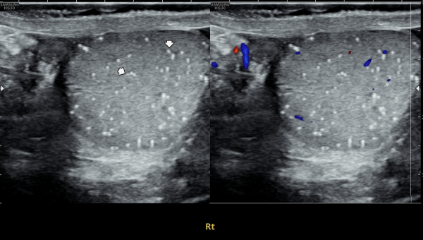
수년전부터 우측 고환의 통증으로 내원 당일 검사한 초음파 사진상 고환의 미석증이 관찰되는 사진입니다.(NIH:24)
The patient presented with right testicular pain for several years.
On the initial ultrasound examination performed on the day of the visit, the right testis shows multiple echogenic foci without acoustic shadowing, consistent with testicular microlithiasis.(NIH:24)
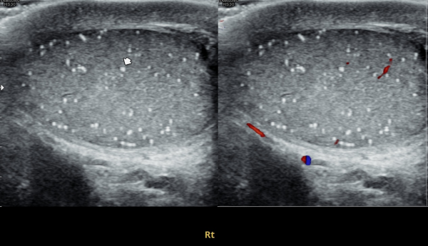
주 2회 14주 동안 정관과 사정관, 정낭 그리고 전립선의 표적 치료후 치료되고 있는 우측 고환 미석증들의 초음파사진입니다.(NIH:13)
This ultrasound image shows improvement of right testicular microlithiasis after targeted treatment of the vas deferens, ejaculatory ducts, seminal vesicles, and prostate.
The treatment was performed twice a week over a period of 14 weeks, and the previously observed tiny calcifications in the testis are gradually improving, suggesting better circulation and recovery of the reproductive tract.(NIH:13)
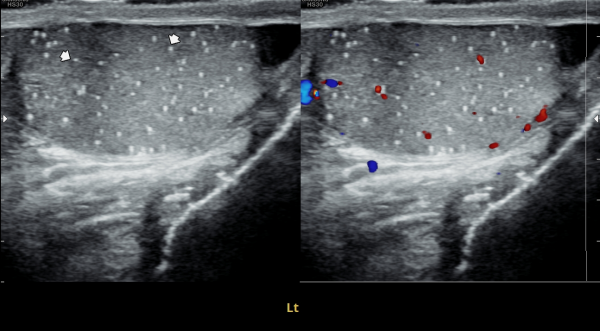
좌측고환의 초음파 검사상 또한 고환의 미석증이 관찰되는 사진입니다.(NIH:24)
On the ultrasound examination of the left testis, multiple tiny echogenic foci without acoustic shadowing are again observed, consistent with testicular microlithiasis.(NIH:24)
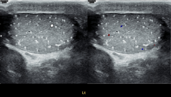
주 2회 14주 동안 정관과 사정관, 정낭 그리고 전립선의 표적 치료후 치료되고 있는 좌측 고환 미석증들의 초음파사진입니다.(NIH:13)
This ultrasound image shows improvement of left testicular microlithiasis after targeted treatment of the vas deferens, ejaculatory ducts, seminal vesicles, and prostate.
The treatment was performed twice a week over a period of 14 weeks, and the previously observed tiny calcifications in the testis are gradually improving, suggesting better circulation and recovery of the reproductive tract.(NIH:13)
These images demonstrate that microlithiasis is present bilaterally, not only on the right side but also within the left testis.
Such findings are characterized by the deposition of microscopic calcifications within the seminiferous tubules.
Clinically, testicular microlithiasis is often asymptomatic but may be associated with chronic testicular pain, infertility, or underlying urogenital conditions, and therefore follow-up and further evaluation may be warranted.
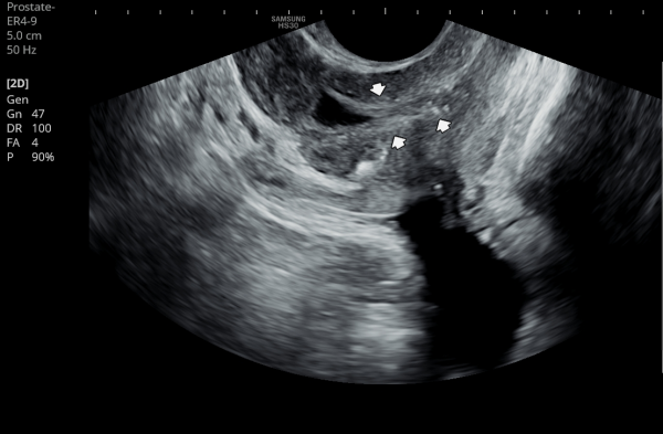
내원 첫 당일 경직장 전립선 초음파 검사상 사정관 입구의 결석들과 사정관 낭종이 관찰되는 사진입니다.(NIH:24)
On the initial transrectal prostate ultrasound, the image shows the presence of calcifications (stones) at the opening of the ejaculatory duct as well as a cystic lesion within the ejaculatory duct (ejaculatory duct cyst).(NIH:24)
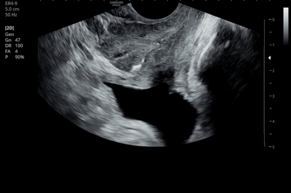
주 2회 14주 동안 전립선과 정낭, 사정관과 정관등의 표적 치료후 사정관의 낭종등이 치료 되고 있는 경직장 전립선 초음파 검사 자료 입니다.(NIH:13)
This transrectal prostate ultrasound image shows improvement of an ejaculatory duct cyst after targeted treatment of the prostate, seminal vesicles, ejaculatory ducts, and vas deferens.
The treatment was performed twice a week over a period of 14 weeks. As a result, the previously noted cyst in the ejaculatory duct is gradually resolving, indicating improved drainage and recovery of normal ductal circulation.(NIH:13)
For the patient, this means that small stones and a cyst are blocking the natural passage where semen normally flows. These findings can explain symptoms such as pelvic pain, difficulty with ejaculation, blood in semen, or infertility.
Treatment may involve addressing these blockages to restore normal flow and relieve associated symptoms.
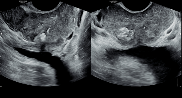
내원 당일 경직정 전립선 정면 사진상 전립선내의 이행구역과 중심 구역 등에 광법위하게 관찰되는 결석과 우측 전립선 부위의 결석이 더 진행된 사진입니다.(NIH:24)
On the transrectal ultrasound image taken on the day of your visit, we can see that multiple stones are widely distributed within the transition zone and central zone of the prostate.
In addition, the image shows that the stones on the right side of the prostate are more advanced and progressed compared with other areas.(NIH:24)
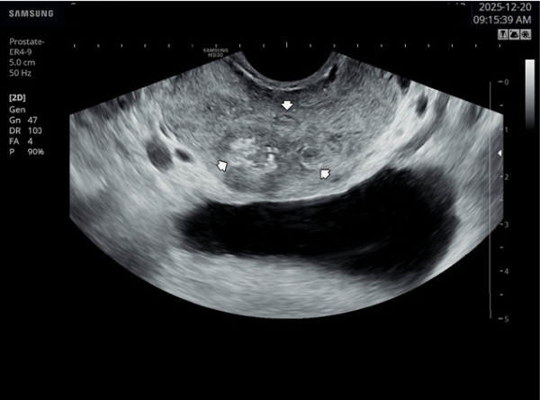
주 2회 14주 동안 전립선과 사정관, 정낭 그리고 정관등의 표적 치료후 전립선의 결석이 치료되고 있는 경직장 전립선 초음파 사진입니다.(NIH:13)
This transrectal prostate ultrasound image shows improvement of prostatic stones after targeted treatment of the prostate, ejaculatory ducts, seminal vesicles, and vas deferens.
The treatment was performed twice a week for 14 weeks, and the previously observed prostatic calcifications are gradually resolving, suggesting improved drainage and recovery of prostate function.(NIH:13)
These images demonstrate that microlithiasis is present bilaterally, not only on the right side but also within the left testis.
Such findings are characterized by the deposition of microscopic calcifications within the seminiferous tubules.
Clinically, testicular microlithiasis is often asymptomatic but may be associated with chronic testicular pain, infertility, or underlying urogenital conditions, and therefore follow-up and further evaluation may be warranted.
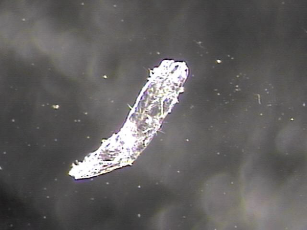
내원 당일 전립선과 사정관 입구와 사정관 그리고 정낭 그리고 정관등의 표적 치료후 치료된 현미경학적 자료 입니다.
This is a microscopic image taken after the targeted treatment of the prostate, ejaculatory duct openings, ejaculatory ducts, seminal vesicles, and vas deferens.
내원 당일 전립선과 사정관 입구와 사정관 그리고 정낭 그리고 정관등의 표적 치료후 치료된 현미경학적 자료 입니다.
This is a microscopic image taken after the targeted treatment of the prostate, ejaculatory duct openings, ejaculatory ducts, seminal vesicles, and vas deferens.
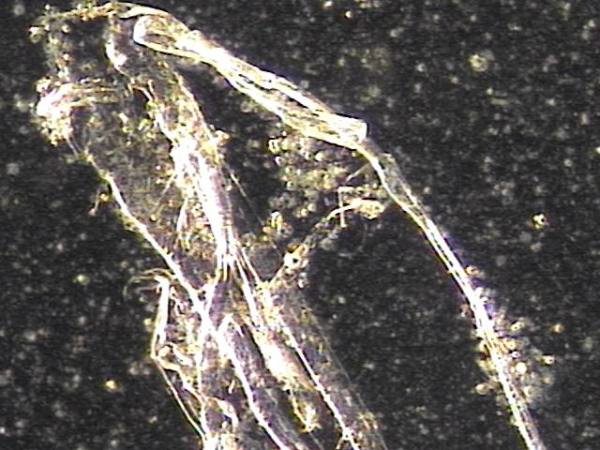
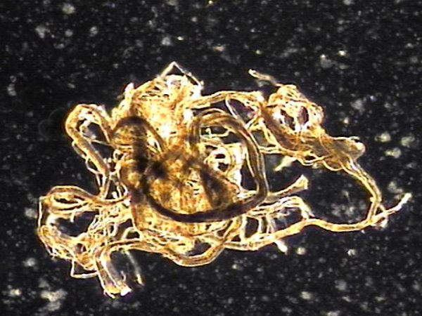
주 2회 전립선과 사정관과 사정관 입구 그리고 정낭과 정낭의 표적 치료후 치료된 현미경학적 자료입니다.
This is a microscopic image obtained after twice-weekly targeted treatment of the prostate, ejaculatory ducts and their openings, seminal vesicles, and vas deferens.
The image shows evidence of treatment effect, including shedding of aged or degenerated epithelial cells that were previously blocking the ducts. These cells are associated with inflammation caused by seminal vesicle contents and prostaglandins.
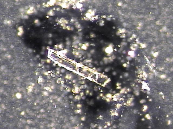
The findings indicate that the treatment helped to clear obstructed and inflamed areas, improving drainage and reducing the blockage that was contributing to symptoms.
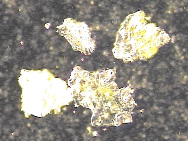
주 2회 전립선과 전립선관, 사정관 입구와 사정관과 정낭 그리고 정관등의 표적 치료후 치료된 현미경학적 자료입니다.
This is a microscopic image taken after twice-weekly targeted treatment of the prostate, prostatic ducts, ejaculatory ducts, seminal vesicles, and vas deferens.
It shows tissue fragments and inflammatory materials that had blocked these passages. Through treatment, these obstructive materials were cleared, helping to improve circulation and function.
This process may support better semen flow and reduce symptoms related to blockage or inflammation.
This microscopic image shows material that was cleared after targeted treatment of the prostate, seminal vesicle, ejaculatory duct, and vas deferens.
The substances include:
- Shed epithelial cells (cells that naturally peeled off the lining)
- Protein-like material and fibrin from past inflammation
- Small inflammatory cells
- Trapped sperm fragments
- Tiny micro-stones (micro-calculi)
These are common findings in conditions such as chronic prostatitis, seminal vesiculitis, or ejaculatory duct inflammation. The treatment helps to remove these blockages and inflammatory materials, allowing for healthier circulation and improved function.
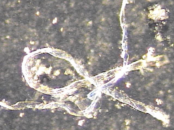
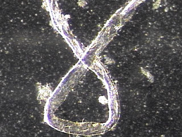
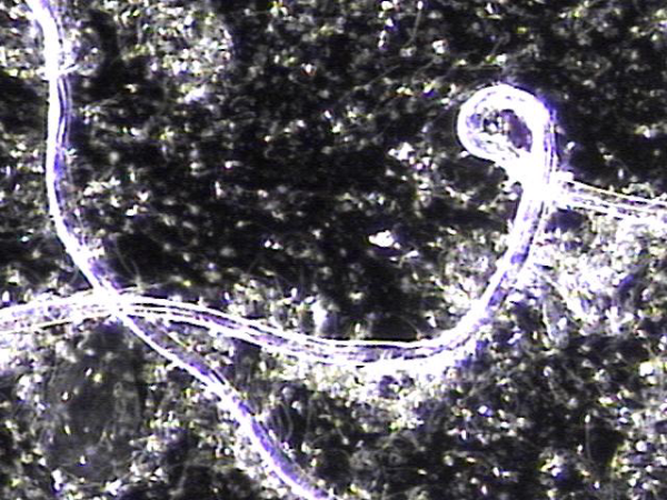
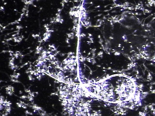
주 2회 전립선과 정낭 그리고 정관등의 표적 치료중 정관을 막고 있는 거짓중층원주상피세포와 사정되지 못한 정자들과 단백질등의 치료된 현미경학적 자료입니다.
This microscopic image was taken after repeated targeted treatment.
It shows that old cells, proteins, and sperm had collected and blocked the vas deferens.
The size of this material confirms that it was stuck inside the vas deferens.
The treatment helped to remove this blockage, allowing the ducts to open and improving the flow of semen.
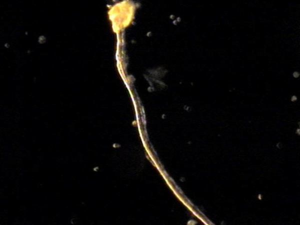
사진 속 현미경 영상은 정관에서 배출된 치료 후 내용물로 보입니다. 관찰되는 소견을 근거로 말씀드리면:
-
길게 뻗은 섬유성 구조와 세포성 잔여물이 함께 보입니다.
-
이는 흔히 노화된 거짓중층원주상피세포, 단백질 덩어리, 또는 정자와 점액질이 엉겨 붙은 물질일 가능성이 높습니다.
-
직경이 비교적 크고 길게 뭉친 형태를 띠기 때문에, 정관 내강을 막아 정액의 흐름을 방해했을 것으로 추정됩니다.
즉, 정관을 막고 있던 주된 원인은
오래된 상피세포 찌꺼기, 단백질 응집물, 그리고 사정되지 못하고 고여 있던 정자들이 엉겨 형성된 덩어리(blockage material) 로 보입니다.
This microscopic image shows material that was removed after targeted treatment of the vas deferens.
The findings suggest that the blockage was mainly caused by:
- Old cells that had shed from the lining,
- Protein debris, and
- Sperm that could not be released and became trapped.
These substances clumped together over time and formed a plug large enough to block the vas deferens, preventing normal flow.
The treatment helped clear this material, allowing for better passage through the duct.
주 2회 전립선과 전립선관, 사정관 입구와 사정관과 정낭 그리고 정관등의 표적 치료후 치료된 현미경학적 자료입니다.
This is a microscopic image taken after twice-weekly targeted treatment of the prostate, prostatic ducts, ejaculatory ducts, seminal vesicles, and vas deferens.
It shows tissue fragments and inflammatory materials that had blocked these passages. Through treatment, these obstructive materials were cleared, helping to improve circulation and function.
This process may support better semen flow and reduce symptoms related to blockage or inflammation.
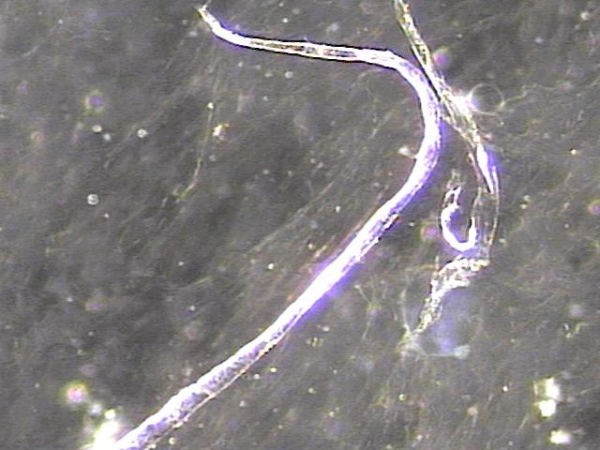
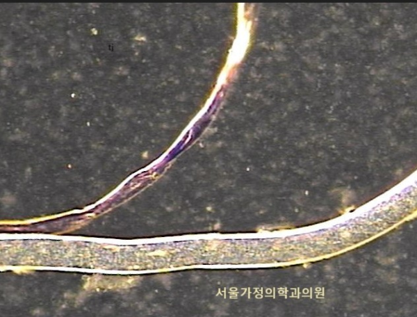
전립선과 정관 그리고 사정관과 정낭등의 표적 치료후 현미경학적 사진과 윗쪽은 정관에 막혀 있던 치료된 사진과 아랫쪽은 전립선관의 치료된 사진.
Here we see two microscopic images after targeted treatment of the prostate, seminal vesicles, ejaculatory ducts, and vas deferens etc.
- The image on the above appears to show material that was once blocking the vas deferens, now cleared after treatment.
- The image on the below seems to show a treated prostatic duct, where the obstruction has also been resolved.
These findings suggest that the therapy helped improve the passageways involved in semen transport.
주 2회 전립선과 전립선관, 사정관 입구와 사정관과 정낭 그리고 정관등의 표적 치료후 치료된 현미경학적 자료입니다.
This is a microscopic image taken after twice-weekly targeted treatment of the prostate, prostatic ducts, ejaculatory ducts, seminal vesicles, and vas deferens.
It shows tissue fragments and inflammatory materials that had blocked these passages. Through treatment, these obstructive materials were cleared, helping to improve circulation and function.
This process may support better semen flow and reduce symptoms related to blockage or inflammation.
주 2회 전립선과 전립선관, 사정관 입구와 사정관과 정낭 그리고 정관등의 표적 치료후 치료된 현미경학적 자료입니다.
These images show cells naturally shed from the prostate and nearby ducts after targeted treatment twice a week, indicating healing and recovery in these areas.
주2회 전립선과 정낭과 사정관 그리고 정관등의 표적 치료후 치료된 상피세포들의 현미경학적 자료입니다.
These images show epithelial cells that have recovered after twice-weekly targeted treatment of the prostate and surrounding ducts.
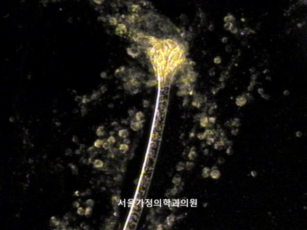
주2회 전립선과 정낭 그리고 정관등의 표적 치료후 치료된 상피 세포의 관으로 탈락된 단백질과 노페물등이 배출되고 있는 현미경학적 사징입니다.
This microscopic image shows the material released after targeted treatments of the prostate, seminal vesicles, and vas deferens performed twice a week.
It demonstrates a small duct-like structure of detached epithelial cells, through which proteins and waste products that had been trapped are now being cleared out as part of the healing process.
주 2회 전립선과 정낭 그리고 정관등의 표적 치료후 치료된 전립선과 사정관, 정낭 그리고 정관등에 막혀 있던 탈락된 상피세포와 프로스타그란딘에 의한
염증세포들이 치료된 현미경학적 사진입니다.
This microscopic image shows the material released after twice-weekly targeted treatments of the prostate, seminal vesicles, and vas deferens.
It includes detached epithelial cells and inflammation-related cells that had been blocked inside the prostate ducts, ejaculatory ducts, seminal vesicles, and vas deferens.
Their release indicates that these areas are opening and clearing as part of the healing process.
주 2회 전립선과 정낭 그리고 정관등의 표적 치료후 치료된 전립선과 사정관, 정낭 그리고 정관등에 막혀 있던 탈락된 상피세포와 단백질과 전해질등 노폐물이 치료돤 현미경학적 사진입니다.
This microscopic image shows the material released after twice-weekly targeted treatments of the prostate, seminal vesicles, and vas deferens.
It includes detached epithelial cells, proteins, electrolytes, and other waste substances that had been trapped inside the prostate ducts, ejaculatory ducts, seminal vesicles, and vas deferens.
Their release indicates that these areas are opening and clearing as part of the healing process.
댓글목록
등록된 댓글이 없습니다.


