전립선자료실
페이지 정보
본문
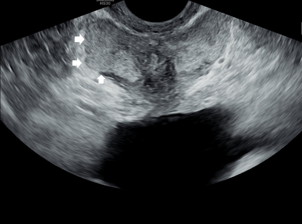
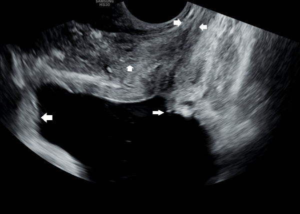
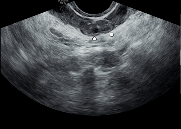
서울가정의학과의원에 첫 내원 당일 지난 5개월동안 회음부 통증과 배뇨장애로 타 비뇨기과 여러곳에서 치료를 했으나 증상의 호전이 없다고 내원 당일 검사한 경직장 전립선 초음파 검사상 사정관 입구의 석회화와 사정관의 섬유화 그리고 정낭의 낭종이 관찰되는 초음파 자료입니다.
On the first visit to Seoul Family Medicine Clinic, the patient reported having perineal pain and urination problems for the past five months, despite receiving treatment at several other urology clinics with no improvement.
A transrectal prostate ultrasound performed on the day of the visit showed calcification at the opening of the ejaculatory duct, fibrosis (scarring) of the duct itself, and cysts in the seminal vesicles.
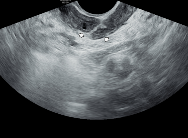
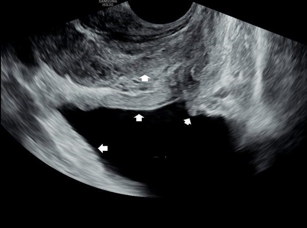
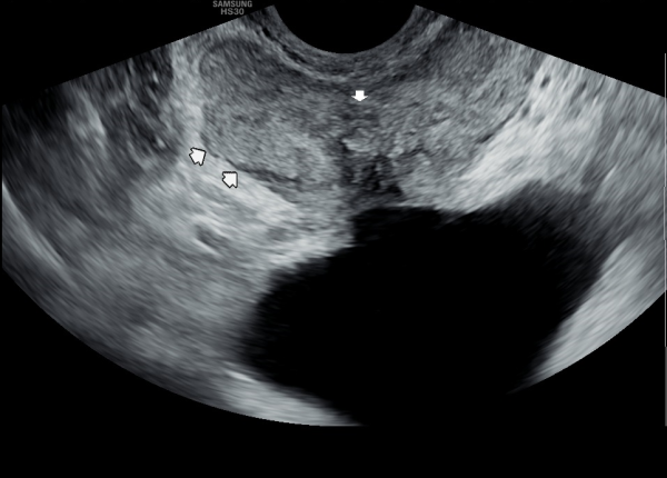
또한 방광벽이 배뇨장애로 두꺼워져 과민성 방광이 의심되는 초음파 사진입니다.
The ultrasound image also shows that the bladder wall has become thickened, likely due to urination difficulties. This may suggest an overactive bladder, which can cause frequent or urgent urination.
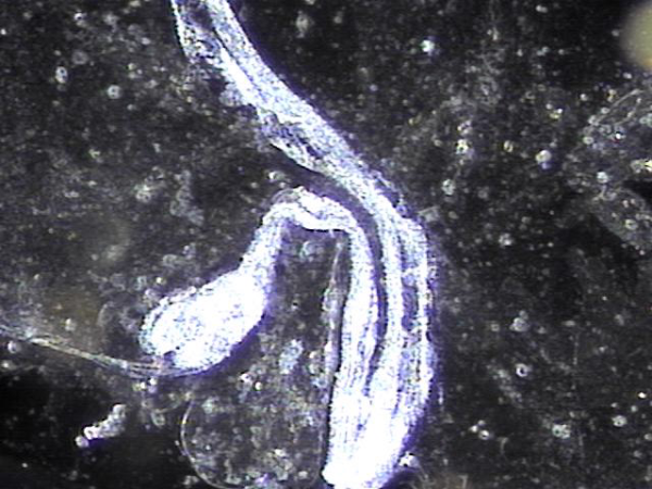
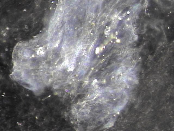
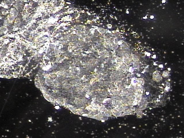
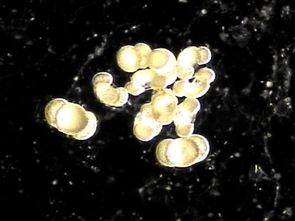
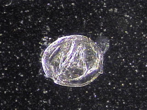
내원 첫날 전립선의 표적 치료후 치료된 정낭의 혈정액과 정자들과 염증들의 현미경학적 자료입니다.
This is a microscopic image taken after your first targeted prostate treatment, showing improvement in the seminal vesicle.
The blood-tinged fluid (hematospermia), sperm, and inflammatory cells have been successfully treated.
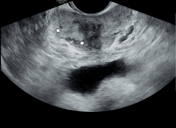
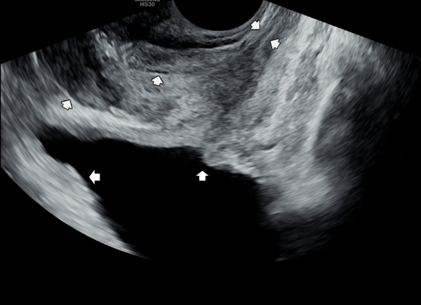
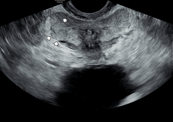
내원 당일 경직장 전립선 초음파 검사상 오랜 세월동안 사정관의 벽이 수명을 다한 거짓 중층 원주 상피 세포가 탈락되어 사정관 입구에 막혀 두텁게 쌓여 있고 요도에도 탈락된 상피 세포가 쌓여서 정낭과 정관 그리고 전립선액 과 배뇨등의 순환 장애를 보이고 있는 경직장 전립선 초음파 사진입니다.
This transrectal prostate ultrasound image, taken on your first visit, shows that over many years, the wall of the ejaculatory duct has been blocked by a buildup of old, shed pseudostratified columnar epithelial cells. These cells have accumulated at the opening of the ejaculatory duct and in the urethra, leading to circulation problems in the seminal vesicles, vas deferens, and prostate fluid, as well as urinary flow issues.
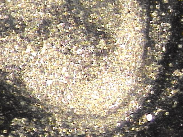
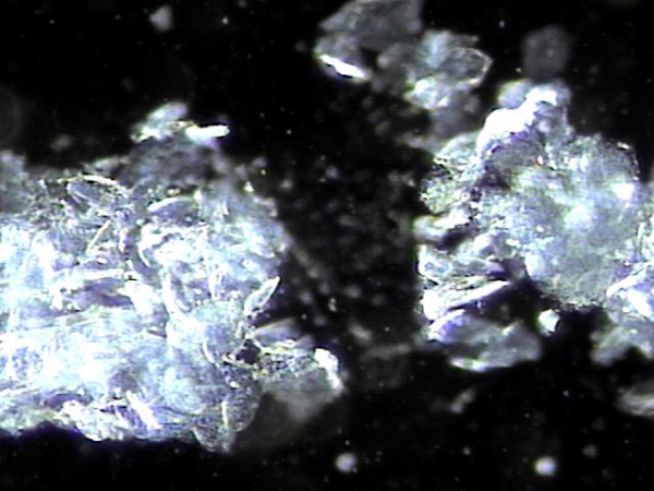
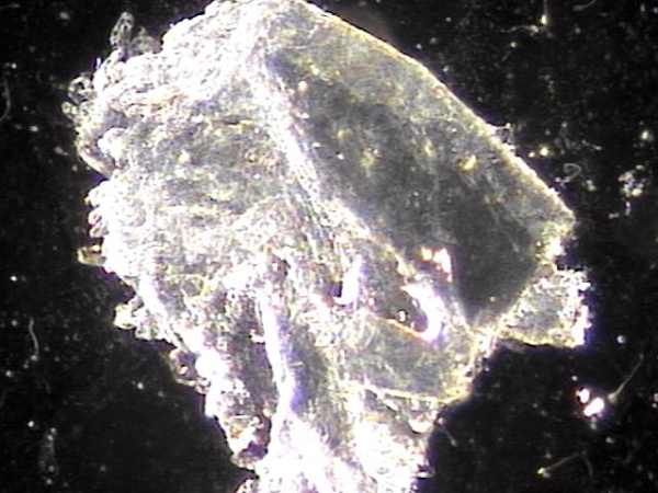
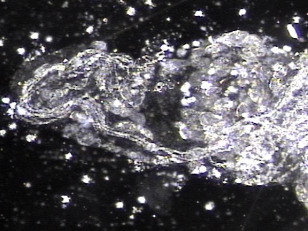
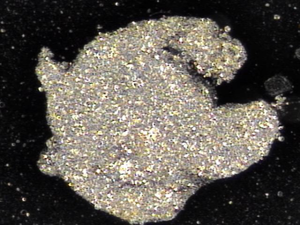
주 2회 전립선의 표적 치료후 사정관 입구에 막혀 있던 탈락된 상피세포와 사정관 벽에 쌓여 있던 상피 세포 덩어리가 치료 된후 관찰한 현미경학적 자료입니다.
This is a microscopic image taken after twice-weekly targeted prostate treatments. It shows the successful removal of shed epithelial cells that had been blocking the opening of the ejaculatory duct, as well as a buildup of epithelial cell clusters that had accumulated along the duct walls.
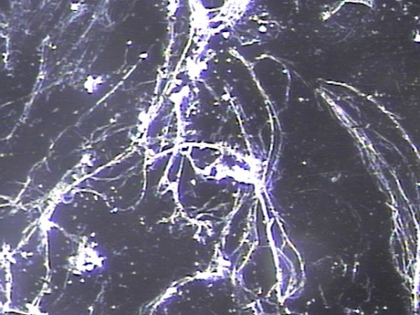
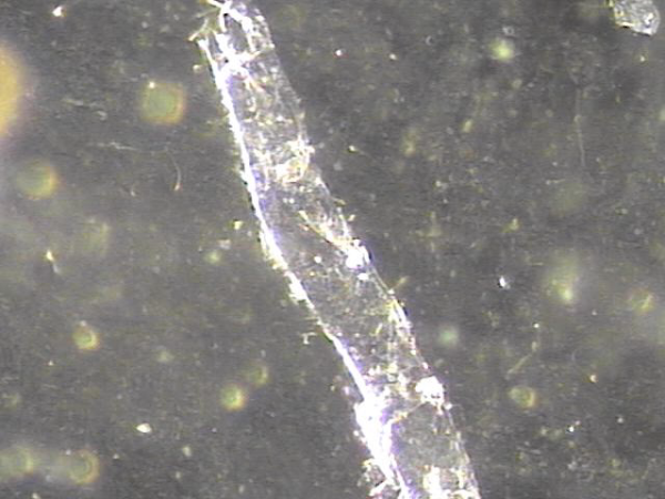
주 2회 전립선의 표적 치료후 전립선관과 사정관 입구와 사정관 내에 쌓여 있던 섬유소 덩어리의 치료된 자료 입니다.
This is post-treatment evidence showing the removal of fibrous clumps that had built up in the prostatic duct, at the entrance of the ejaculatory duct, and within the ejaculatory duct itself, following twice-weekly targeted prostate treatments.
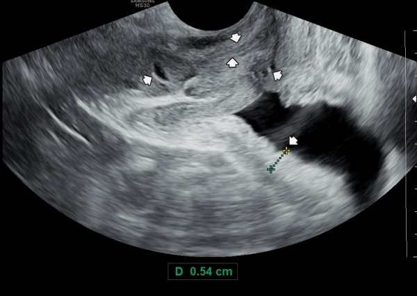
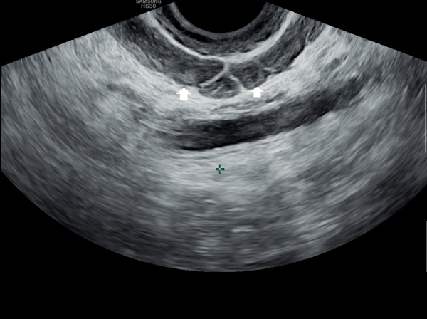
내원 첫날 정면 경직장 전립선 초음파 정낭의 검사상 사정관 입구의 순환 장애로 정낭의 낭종이 관찰되는 초음파 사진입니다.
This is a transrectal prostate ultrasound image taken on the first visit, showing a cyst in the seminal vesicle caused by a circulation blockage at the entrance of the ejaculatory duct.
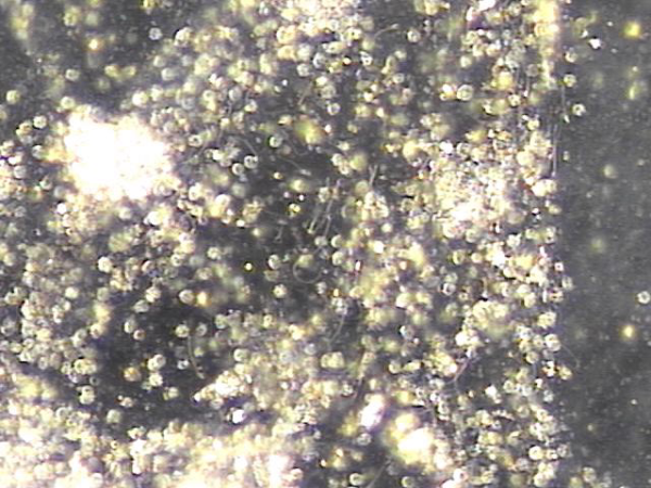
주 2회 전립선의 표적 치료후 치료된 정낭의 혈정액들의 현미경학적 자료입니다.
After receiving targeted prostate treatment twice a week, this microscopic image shows the improvement in the seminal vesicles,
where the blood in the semen (hematospermia) has been treated.
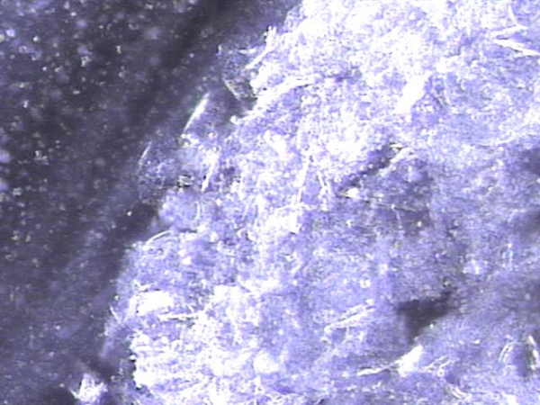
주 2회 전립선의 표적 치료후 전립선관과 사정관 입구와 사정관 내에 쌓여 있던 섬유소 덩어리의 치료된 자료 입니다.
This is post-treatment evidence showing the removal of fibrous clumps that had built up in the prostatic duct, at the entrance of the ejaculatory duct, and within the ejaculatory duct itself, following twice-weekly targeted prostate treatments.
전립선의 경직장 전립선 초음파상 우측 전립선의 순환장애로 전립선의 이행구역내 탈락된 상피세포들이 쌓여 결절을 형성하고 전립선액 즉 구연산과 섬유소용해소 그리고 산성포스파타제등의 순환 장애로 전립선의 낭종이
관찰되는 사진입니다.
This transrectal prostate ultrasound image shows that, due to impaired circulation on the right side of the prostate, exfoliated epithelial cells have accumulated in the transition zone, forming a nodule. As a result of poor circulation of prostatic fluid—such as citrate, fibrinolysin, and acid phosphatase—prostatic cysts are also observed.
주 2회 전립선의 표적 치료후 사정관 입구에 막혀 있던 탈락된 상피세포와 사정관 벽에 쌓여 있던 상피 세포 덩어리가 치료 된후
관찰한 현미경학적 자료입니다.
This is a microscopic image taken after twice-weekly targeted prostate treatments. It shows the successful removal of shed epithelial cells that had been blocking the opening of the ejaculatory duct, as well as a buildup of epithelial cell clusters that had accumulated along the duct walls.
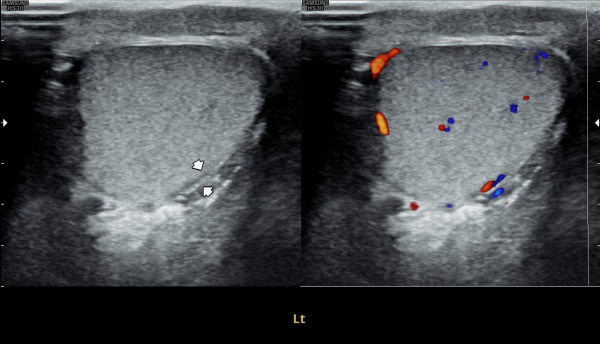
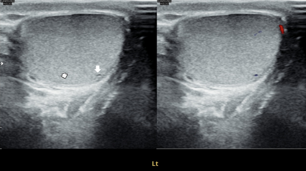
첫 내원 당일 검사한 고환의 초음파 사진상 앉아서 생활하는 직업상 고환 동맥의 순환 장애로 죽상동맥경화증의 소견을 보여 식이요법과 운동요법을 말씀드리고 정관의 표적 치료를 시작한 사진입니다.
On the day of your first visit, the testicular ultrasound showed signs of reduced blood flow in the testicular artery, likely from long hours of sitting. This may have caused early signs of atherosclerosis. We discussed healthy lifestyle changes, including diet and exercise, and started targeted treatment for the vas deferens.
주 2 회 전립선과 정낭 그리고 정관등에 표적 치료후 막혀 있던 거짓 중층 원주 상피 세포의 치료된 현미경학적 자료입니다.
After receiving targeted treatment twice a week to the prostate, seminal vesicles, and vas deferens, this microscopic image shows the cleared-out buildup of aging pseudostratified columnar cells that had been causing blockages.
4개월 가량 주 2회 전립선과 정낭 그리고 사정관과 정관등의 표적 치료후 사정관 주위에 막혀 있던 탈락된 상피 세포 덩어리가 치료되고 정액의 순환장애가 관찰되는 추적 경직장 전립선의 초음파 사진입니다.
This is a follow-up transrectal ultrasound image of the prostate, taken after approximately four months of twice-weekly targeted treatment of the prostate, seminal vesicles, ejaculatory ducts, and vas deferens. It shows that the clumps of shed epithelial cells blocking the area around the ejaculatory ducts have been cleared, although some signs of impaired semen flow are still visible.
주 2회 전립선과 사정관과 정낭 그리고 정관의 표적 치료후 전립선관내와 사정관내 그리고 정관내 등에 막혀 있던 오래된 상피 세포 덩어리가 치료된 현미경학적 사진입니다.
This is a microscopic image taken after twice-weekly targeted treatment of the prostate, ejaculatory ducts, seminal vesicles, and vas deferens. It shows that old clumps of accumulated epithelial cells, which were blocking the prostate ducts, ejaculatory ducts, and vas deferens, have been successfully cleared.
4개월 가량 주 2회 정낭의 표적 치료후 정낭의 낭종들이 감소하고 순환되는 추적 경직장 전립선 초음파 사진입니다.
This is a follow-up transrectal prostate ultrasound image taken after approximately four months of twice-weekly targeted treatment of the seminal vesicles. The image shows that the cysts in the seminal vesicles have decreased and circulation has improved.
주 2회 사정관과 정낭의 표적 치료후 치료된 상피세포 덩어리와 막혀 순환장애로 고여 있던 정자들의 현미경학적 자료입니다.
This is a microscopic image taken after twice-weekly targeted treatment of the ejaculatory ducts and seminal vesicles. It shows the removal of accumulated epithelial cell clusters and stagnant sperm that had been trapped due to circulation blockage.
오렌 세월 동안 전립선의 이행 구역의 순환 장애로 전립선 결절들이 관찰되는 경직장 전립선 초음파 사진입니다.
This transrectal ultrasound image shows nodules in the prostate transition zone, which appear to have developed over a long period due to impaired circulation in that area.
오래된 전립선의 결절이 표적 치료후 배출된 섬유소덩어리의 현미경학적 자료입니다.
This is a microscopic image of fibrous clumps that were released after targeted treatment of long-standing prostate nodules.
추적 경직장 전립선 초음파 사진상 사정관 주위에 탈락되어 막혀 있던 상피 세포가 현저히 감소하고
과민성 방광을 생기게 했던 두터운 방광벽이 감소하고 있는 사진입니다.
"This follow-up transrectal prostate ultrasound image shows a significant reduction in the accumulated exfoliated epithelial cells that had previously obstructed the area around the ejaculatory ducts, along with a noticeable thinning of the previously thickened bladder wall,
which had contributed to overactive bladder symptoms.
4개월 동안 정관의 표적 치료와 직장 생활중 장시간 앉은 업무를 개선하여 운동을 겸한후 추적 고환의 초음파 사진장
고환동맥의 축상경화증 소견이 치료되고 있는 사진입니다.
This follow-up scrotal ultrasound image, taken after four months of targeted treatment for the vas deferens and lifestyle improvements including reduced prolonged sitting during office work and regular exercise, shows improvement in axial arteriosclerosis of the testicular artery.
8개월 주2회 표적 치료후 추적 경직장 전립선 초음파 사진상 사정관 주위에 탈락되어 막혀 있던 상피 세포가 현저히 감소하고
과민성 방광을 생기게 했던 두터운 방광벽이 감소하고 있는 사진입니다.
"This follow-up transrectal prostate ultrasound image shows a significant reduction in the accumulated exfoliated epithelial cells that had previously obstructed the area around the ejaculatory ducts, along with a noticeable thinning of the previously thickened bladder wall,
which had contributed to overactive bladder symptoms.
전립선의 이행구역 부위에 쌓인 탈락된 상피 세포들이 결절을 형성하여 전립선의 순환 장애와 회음부 압박등의 만성 골반통증후군의 증상을 일으키는 원인중 하나를 치료키 위해 직장 점막과 점막하층 그리고 근육층과 장간막등에 집중적 표적 치료로 직장벽의 손상으로 출혈을 일으킬수 있으나 치료후
배출된 상피세포 덩어리의 현미경학적 자료입니다.
This is a microscopic image showing clumps of epithelial cells that were released after targeted treatment. These cells had accumulated in the transitional zone of the prostate, forming nodules that may have caused poor circulation, pressure in the perineum, and symptoms of chronic pelvic pain syndrome.
To treat this, targeted therapy was applied to the rectal mucosa, submucosa, muscle layer, and surrounding tissues. While this focused treatment may sometimes cause temporary bleeding due to rectal wall irritation, it helps release the obstructing epithelial cells and improve circulation and symptoms.
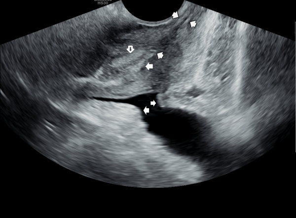
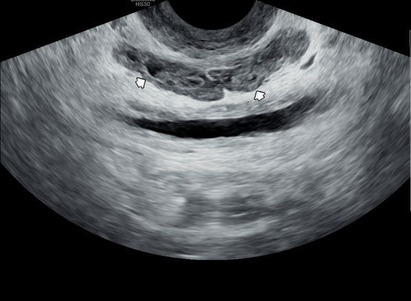
주 2회 전립선과 사정관과 정관 그리고 정낭 등의 표적 치료후 정낭 낭종들이 치료된 경직장 전립선 초음파 사진입니다.
This is a transrectal prostate ultrasound image showing that, after targeted treatment of the prostate, ejaculatory ducts, vas deferens, and seminal vesicles twice a week, the seminal vesicle cysts have been successfully treated.
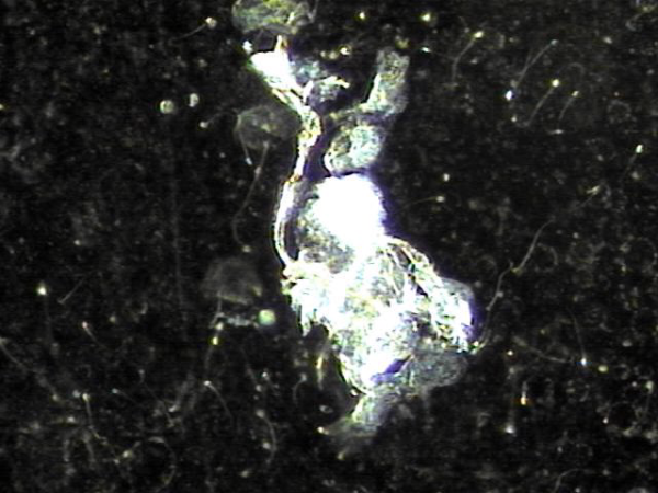
전립선의 표적 치료중 탈락되어 오래되고 사정관과 정관, 전립선관과 전립선관을 막고 있는 탈락된 상피세포들의 치료된 현미경학적 사진입니다.
This is a transrectal prostate ultrasound image taken after twice-weekly targeted treatments of the prostate, ejaculatory ducts, vas deferens, and seminal vesicles. It shows that the seminal vesicle cysts have been successfully treated.
6개월 가량 주 2회 전립선의 표적 치료를 했으나 탈락된 상피세포가 장기간 축적되어 전립선의 결절과 비대증을 일으키고 있는
경직장 전립선 초음파 사진입니다.
This transrectal prostate ultrasound image shows nodular formation and enlargement of the prostate caused by long-term accumulation of shed epithelial cells, despite approximately six months of twice-weekly targeted prostate treatment.
주 2회 전립선의 표적 치료후 전립선과 정낭과 사정관 그리고 정관등의 상피세포들이 수명을 다한후 탈락되어
오랜 세월 막혀 순환 장애를 일으킨 상피 세포들의 현미경학적 자료입니다.
This microscopic image shows shed epithelial cell clusters that had accumulated over many years and caused circulation obstruction in the prostate, seminal vesicles, ejaculatory ducts, and vas deferens. These cells were released following twice-weekly targeted prostate treatments.
8개월 주2회 전립선의 표적 치료후 전립선의 이행구역에 생긴 결절과 비대가 감소하고 있는 경직장 전립선 초음파 사진입니다.
This transrectal prostate ultrasound image shows that, after 8 months of twice-weekly targeted prostate treatment, the nodules and enlargement in the transitional zone of the prostate have been reduced.
8개월 주 2회 정관의 표적 치료후 정관의 순환 장애를 일으키고 있는 결석들의 현미경학적 자료입니다.
This microscopic image shows calcifications in the vas deferens that have been causing circulation issues, observed after 8 months of twice-weekly targeted treatment of the vas deferens.
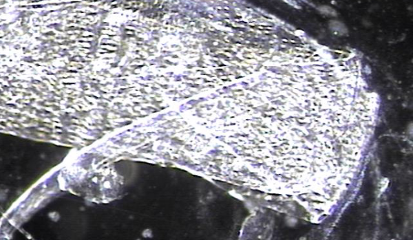
전립선의 주변 구역에 커지고 오래된 상피 세포 덩어리가 표적 치료후 치료된 현미경 학적 자료입니다.
This microscopic image shows an old and enlarged cluster of epithelial cells in the peripheral zone of the prostate
that has been treated following targeted therapy.
주 2회 정관과 사정관등의 표적 치료 중 일반 운동과 전립선에 탁월한 효과를 보이는 PST exercise를 겸해서 치료후 첫 내원시 고환의 죽상 동맥 경화증이
치료된 고환의 초음파 사진입니다.
This is a follow-up scrotal ultrasound image taken during the initial visit after combining targeted treatment of the vas deferens and ejaculatory ducts (twice a week) with general exercise and PST exercises, which are highly effective for prostate health. The image shows improvement in testicular atherosclerosis.
주 2회 지속적으로 치료중 전립선관과 사정관 그리고 정관등에 막혀 있던 탈락된 상피 세포 덩어리의 현미경 학적 사진입니다.
This is a microscopic image showing clumps of shed epithelial cells that had been blocking the prostatic ducts, ejaculatory ducts,
and vas deferens during ongoing targeted treatment twice a week.
주 2회 전립선과 정낭 그리고 정관등의 표적 치료중 주로 정관에 막혀 있던 성분들의 치료된 현미경 학적 자료입니다.
This is microscopic data of treated components, mainly those obstructing the vas deferens, during targeted therapy of the prostate,
seminal vesicles, and vas deferens performed twice weekly.
주 2회 전립선과 사정관, 정낭 그리고 정관등의 표적 치료를 했으나 추적 정낭과 전립선 등의 검사상 치료된 정낭의 낭종들이 다시 관찰되어
주 3회 이상 전립선의 표적 치료가 필요하다고 요구되는 정낭의 추적 경직장 초음파 사진입니다.
This is a follow-up transrectal ultrasound image of the seminal vesicles and prostate after twice-weekly targeted treatments of the prostate, ejaculatory ducts, seminal vesicles, and vas deferens. Although improvements were seen, some treated seminal vesicle cysts are still visible. Based on this, continuing treatment three times a week or more is recommended for better recovery.
주 2회 전립선의 표적 치료후 이행구역 전립선의 결절들의 크기가 감소하고 사정관 입구에 막혀있던 탈락된 상피 세포가 치료되고 있는 추적 경직장 전립선
초음파 사진입니다.
This is a follow-up transrectal prostate ultrasound image taken after twice-weekly targeted prostate therapy, showing a reduction in the size of nodules in the transitional zone and ongoing resolution of detached epithelial cell blockage at the ejaculatory duct openings.
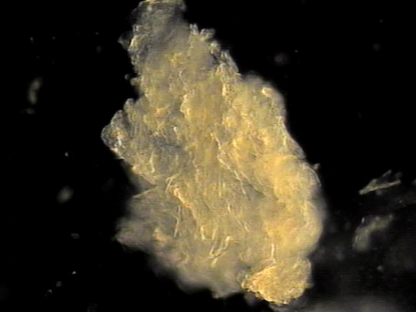
전립선과 사정관,정낭 그리고 정관등에서 치료된 상피세포 덩어리의 현미경학적 자료입니다.
Here is microscopic evidence of epithelial cell clusters that have been cleared following targeted therapy of the prostate, ejaculatory ducts, seminal vesicles, and vas deferens.
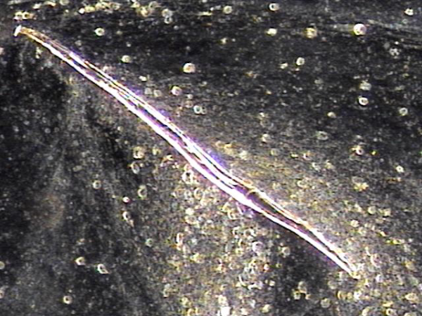
반드시 치료 되어야할 전립선관내 그리고 사정관과 정관등에 막혀 있던 상피세포덩어리가 치료된 현미경학적 자료입니다.
Here is microscopic evidence showing the successful removal of epithelial cell clusters that had been obstructing the prostatic ducts, ejaculatory ducts, and vas deferens—areas that require proper treatment.
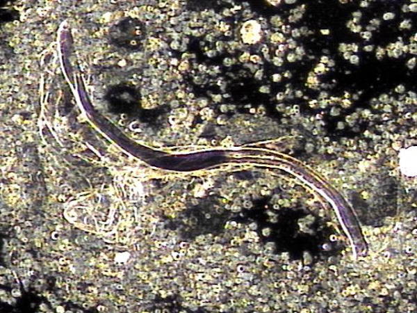
전립선과 정낭, 사정관과 정관등의 순환 장애를 일으키는 압축된 상피세포와 프로스타그란딘으로 막혀있던 염증 세포가 치료된 현미경학적 자료입니다.
Here is microscopic evidence showing the resolution of compressed epithelial cells and inflammatory cells, previously blocked by prostaglandins and causing circulation disturbances in the prostate, seminal vesicles, ejaculatory ducts, and vas deferens.
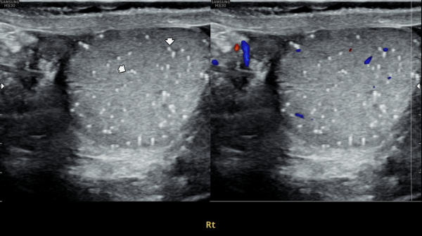
고환의 통증으로 내원한 환자분의 고환의 초음파 검사상 우측 고환의 미석증이 관찰되는 초음파 사진입니다.
Here is an ultrasound image of a patient who presented with testicular pain, showing microlithiasis in the right testis.
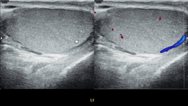
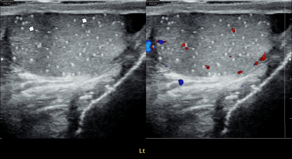
고환의 통증으로 내원한 환자분의 고환의 초음파 검사상 좌측 고환의 미석증이 관찰되는 초음파 사진입니다.
Here is an ultrasound image of a patient who presented with testicular pain, showing microlithiasis in the left testis.
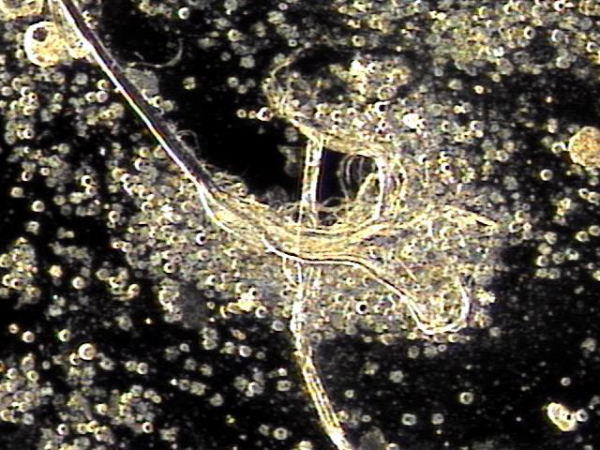
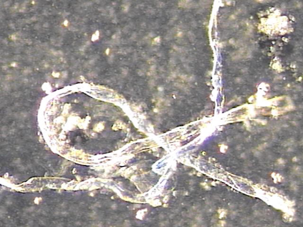
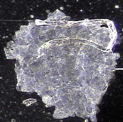
정관을 막아 고환의 미석증을 일으컸던 상피세포가 치료된 현미경학적 자료입니다.
댓글목록
등록된 댓글이 없습니다.


