전립선자료실
페이지 정보
본문
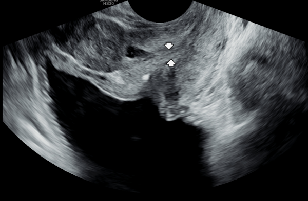
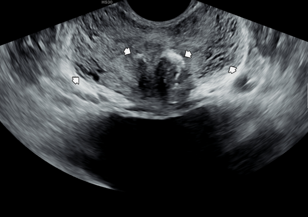
수년전부터 회음부에 통증이 있고 빈뇨가 심하다고 내원 당일 검사한 경직장 전립선 초음파 사진상 사정관의 낭종과 사정관이 좁아지고 있는 자료입니다.
A transrectal prostate ultrasound image taken on the day of the visit shows a cyst in the ejaculatory duct
and narrowing of the duct in a patient who had been experiencing perineal pain and severe urinary frequency for several years.
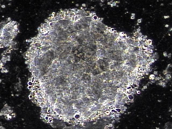
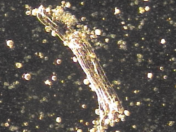
전립선의 표적 치료중 사정관, 정관 그리고 전립선관등에 막혀 순환 장애을 일으키는 탈락된 상피 세포 덩어리의 현미경학적 자료입니다.
This is a microscopic image showing clusters of shed epithelial cells that were blocking the ejaculatory ducts, vas deferens, and prostatic ducts,
leading to circulation problems. These findings were observed during targeted prostate therapy.
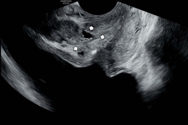
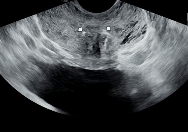
5년뒤 바쁜 일상 생활로 전립선의 관리를 하지 못하고 고환의 통증과 빈뇨 그리고 회음부 통증이 심해진다고 내원 당일 검사한 경직장 전립선 초음파 검사상 사정관 낭종과 낭종내 미세 결석이 생기고 사정관이 탈락된 상피 세포가 70%가량 좁아지고 전립선관도 순환 장애로 전립선 낭종이 관찰되는 초음파 사진입니다.
This transrectal prostate ultrasound image was taken on the day of the visit, five years later. Due to a busy lifestyle, the patient was unable to maintain prostate care and began experiencing worsening testicular pain, frequent urination, and perineal discomfort. The scan shows an ejaculatory duct cyst with microcalcifications inside, as well as narrowing of about 70% of the duct caused by accumulated epithelial cells. Circulatory issues have also led to the formation of prostatic cysts in the prostatic ducts.
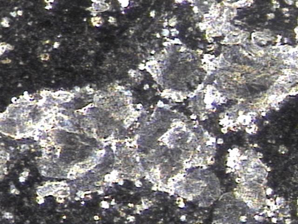
주 2~3회 전립선의 표적 치료후 사정관과 전립선관 그리고 정관등에 순환 장애를 일으킨 탈락된 상피 세포덩어리와 프로스타그란딘등에 의한 염증 세포들이 치료된 현미경 학적 자료입니다.
This is a microscopic image taken after receiving targeted prostate therapy 2 to 3 times per week. It shows that clusters of shed epithelial cells, which had been blocking circulation in the ejaculatory ducts, prostatic ducts, and vas deferens, along with inflammatory cells caused by substances like prostaglandins, have been successfully treated.
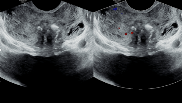
5년전 내원 당일 검사한 경직장 전립선 초음파 사진상 좌우 사정관 입구의 막혀있는 미세 결석들과 전립선관의 순환장애로 전립선의 낭종들이 관찰되는 사진입니다.
A transrectal prostate ultrasound image taken on the day of the initial visit five years ago shows microcalcifications blocking the openings of both ejaculatory ducts, as well as prostatic cysts caused by impaired circulation in the prostatic ducts.
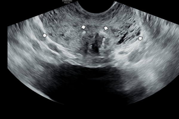
전립선과 정낭, 정관 그리고 망울요도샘등의 표적 치료후 치료된 상피세포 덩어리의 현미경학적 자료입니다.
This is a microscopic image showing clusters of epithelial cells that were successfully treated and cleared after targeted therapy to the prostate, seminal vesicles, vas deferens, and bulbourethral glands.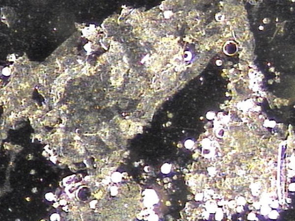
5년뒤 바쁜 일상 생활로 상기 환자분이 적절히 치료를 하지 않고 고환의 통증과 사정 장애와 사정통 그리고 회음부의 통증이 심하다고 내원하여 추적 검사한 경직장 전립선 초음파 검사상 좌우 사정관입구의 결석이 커지고
전립선의 다발성 낭종들이 만들어지고 전립선이 비대해 지고 있는 사진입니다.
Five years later, the patient returned with complaints of worsening testicular pain, ejaculation difficulties, pain during ejaculation, and perineal discomfort, having not received appropriate treatment due to a busy lifestyle. A follow-up transrectal prostate ultrasound showed enlarged stones at the openings of both ejaculatory ducts, multiple new cysts forming in the prostate, and progressive prostate enlargement.
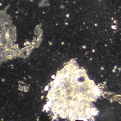
전립선의 표적 치료후 치료된 상피세포 덩어리의 현미경학적 사진입니다.
A microscopic image of treated epithelial cell clusters following targeted prostate therapy.
5년전 경직장 전립선 초음파 사진상 좌우 사정관 입구의 결석과 좌측 전립선의 주변구역의 낭종이 관찰되는 초음파 사진입니다.
A transrectal prostate ultrasound image from five years ago shows calcifications at the openings of both ejaculatory ducts and a cyst in the peripheral zone of the left prostate.
상기 환자분의 5년뒤 검사한 추적 경직장 전립선의 초음파 검사상 좌우 사정관 입구의 결석이 커지고 좌측 전립선의 주변구역의 전립선 낭종들이 순환장애로 커지고 많아지고 비대해지고 있는 추적 초음파 사진입니다.
This follow-up transrectal prostate ultrasound, taken five years later, shows that the stones at the openings of both ejaculatory ducts have increased in size. In addition, multiple cysts in the peripheral zone of the left prostate have become larger and more numerous due to circulation issues, and the prostate itself is gradually enlarging.
주 2회 전립선의 표적 치료후 탈락된 거짓중층 원주 상피 세포 덩어리의 현미경학적 자료입니다.
This is a microscopic image of detached pseudostratified columnar epithelial cell clusters following targeted prostate therapy twice a week.
탈락된 정관의 상피 세포가 순환 장애를 일으켜 고환의 섬유화를 일으키고 있는 고환의 초음파 사진입니다.
This is an ultrasound image of the testis showing fibrosis caused by impaired circulation due to detached epithelial cells from the vas deferens.
전립선의 표적 치료후 전립선과 사정관, 정낭 그리고 정관등에 막혀 있던 상피 세포 덩어리와 염증 세포들의 현미경학적 자료입니다.
This is a microscopic image taken after targeted prostate therapy, showing clusters of epithelial cells and inflammatory cells that had been blocking the prostate, ejaculatory ducts, seminal vesicles, and vas deferens.
수년간 전립선의 표적 치료를 받지 않을 경우 탈락된 정관의 상피 세포로 고환의 섬유화가 점점 진행되고 있는 고환의 추적 초음파 사진입니다.
This is a follow-up ultrasound image of the testis showing gradually progressing fibrosis, likely caused by detached epithelial cells from the vas deferens,
in a case where targeted prostate treatment was not received for several years.
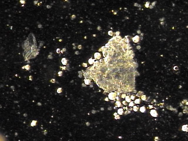
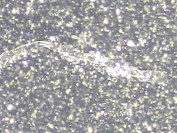
정관의 표적 치료후 치료된 정관 결석의 현미경학적 사진입니다.
This is a microscopic image of a treated vas deferens stone following targeted therapy.
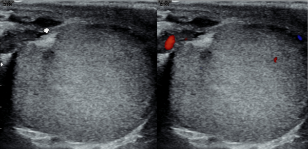
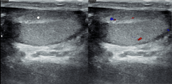
지금부터 5년전 고환의 초음파 검사상 고환의 섬유화가 시작되는 사진입니다.
This is an ultrasound image of the testis from 5 years ago, showing the early signs of testicular fibrosis.
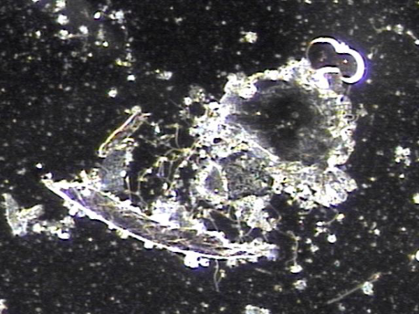
전립선과 정낭 그리고 사정관과 정관등에 막혀 있던 상피 세포의 치료된 현미경학적 사진입니다.
This is a microscope image showing treated cell buildup that had been blocking areas like the prostate, seminal vesicles, ejaculatory ducts, and vas deferens.
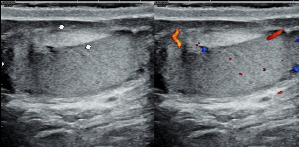
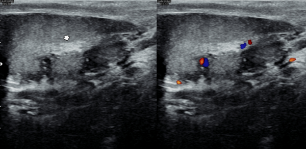
상기 고환의 초음파 검사후 치료를 받지 않고 고환의 통증과 사정장애등으로 내원하여 추적 검사한 초음파 사진상 고환의 섬유화가 점점 진행되는 사진입니다.
This is a follow-up ultrasound image showing progressive testicular fibrosis. The patient returned with testicular pain and ejaculation difficulties after not receiving treatment following the previous ultrasound.
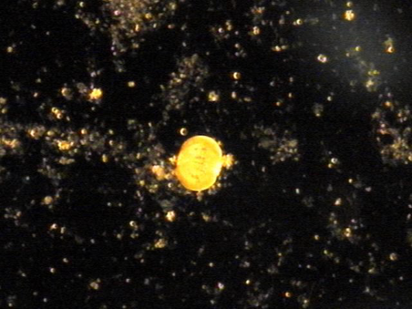
주 2회 전립선의 표적 치료후 전립선관과 사정관 그리고 정관 등에 막현 탈락된 상피 세포 덩어리와 사정되지 못한 정자들의 현미경학적 사진입니다.
This is a microscopic image taken after twice-weekly targeted prostate treatments. It shows clumps of old, shed epithelial cells and sperm
that were unable to be ejaculated, which had been blocking the prostate ducts, ejaculatory ducts, and vas deferens.
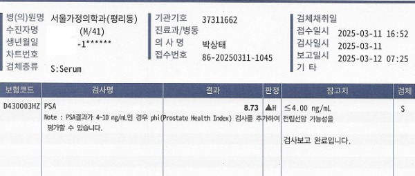
수주전부터 하복부의 통증과 배뇨시 불쾌감 그리고 사정시 혈정액을 호소하면서 내원하신 분의 임상 병리에 의래한 혈액학적 PSA 검사상 8.73 ng/mL가 높게 나온 자료입니다.
This patient came to our clinic after experiencing lower abdominal pain, discomfort during urination, and blood-tinged semen during ejaculation
for several weeks. A blood test (PSA test) showed an elevated PSA level of 8.73 ng/mL, which is higher than the normal range.
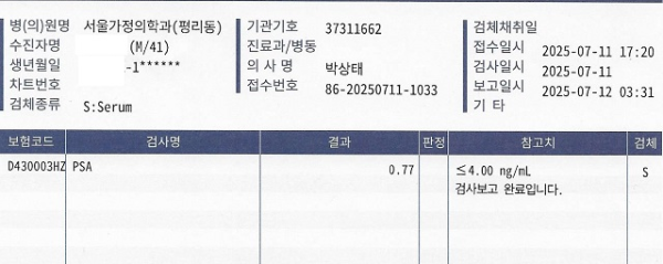
4개월동안 주 2회 전립선과 사정관, 정낭 그리고 정관 등의 표적 치료후 혈액학적 PSA 추적 검사상 0.77 ng/mL로 정상 범위로 치료된 자료입니다.
After receiving targeted treatment for the prostate, seminal vesicles, ejaculatory ducts, and vas deferens twice a week for four months,
the follow-up blood test showed a PSA level of 0.77 ng/mL, which is within the normal range.
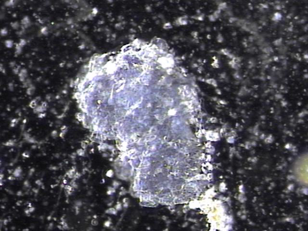
4개월 동안 주2회 전립선의 표적 치료중 전립선관과 사정관, 정낭 그리고 정관등에서 막혀서 PSA 수치를 높인 탈락된 막혀 있던 상피 세포 덩어리의
치료된 현미경학적 자료입니다.
This is a microscopic image of the treated clumps of shed epithelial cells that were blocking the prostate ducts, ejaculatory ducts, seminal vesicles, and vas deferens. These blockages had contributed to an elevated PSA level, but after four months of targeted prostate treatment twice a week,
the blockages were successfully cleared.
댓글목록
등록된 댓글이 없습니다.


