전립선자료실
페이지 정보
본문
17세 고등학교 2학년 남성이 한달전부터 사정시 혈정액이 나오고 관절과 근육통으로 인근 비뇨기과에서 치료를 했으나 증상의 호전이 없다고
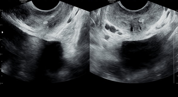
내원 당일 경직장 전립선 초음파 검사상 좌측 사정관 입구에 결석이 관찰되는 측면과 정면 초음파 사진입니다.
A 17-year-old male high school junior presented with hematospermia and joint and muscle pain for the past month. Despite treatment at a local urology clinic, there was no improvement in symptoms. On the day of his visit, transrectal prostate ultrasonography revealed a stone at the entrance of the left ejaculatory duct.
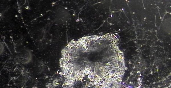
전립선과 사정관, 정관 그리고 정낭등의 표적치료후 치료된 현미경학적 자료입니다.
This is microscopic evidence of successful treatment following targeted therapy of the prostate, ejaculatory ducts, vas deferens, and seminal vesicles.
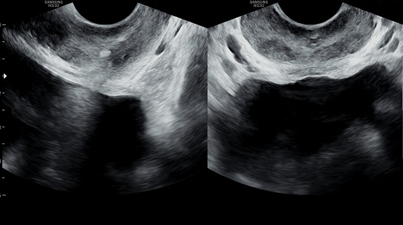
내원 당일 검사한 경직장 전립선 초음파 검사상 사정관 결석과 정낭의 낭종들이 관찰되는 초음파 사진입니다.
On the day of the initial visit, transrectal prostate ultrasound revealed ejaculatory duct stones and multiple seminal vesicle cysts.
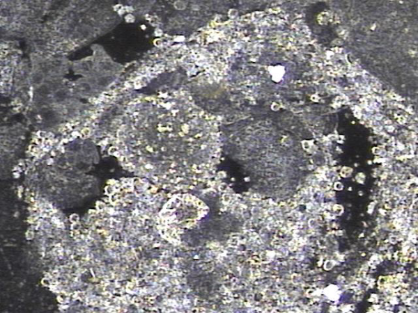
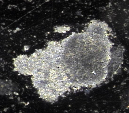
첫 내원일후 한달 보름 주 2~3회 전립선의 표적 치료후 사정관과 정낭 그리고 사정관입구에 막혀 있던 탈락된 상피 세포가 치료된 현미경학적 자료입니다.
This is a microscopic image showing the improvement after 6 weeks of targeted prostate treatment, performed 2 to 3 times a week. The treatment helped clear away the accumulated, shed epithelial cells that had been blocking the ejaculatory ducts, seminal vesicles, and the opening of the ejaculatory ducts.
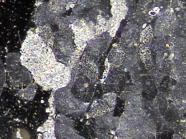
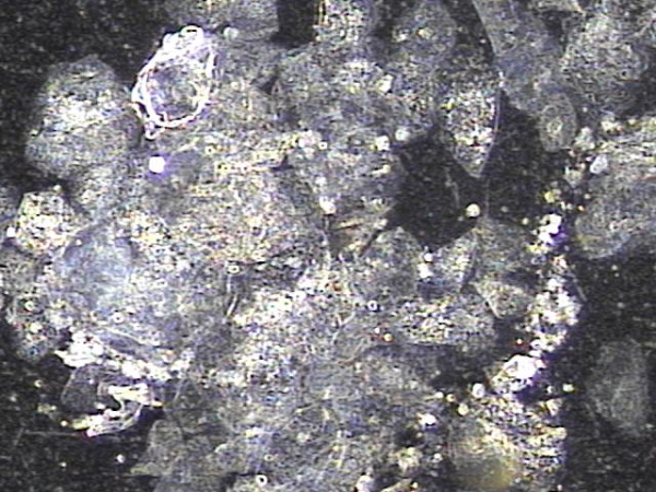
전립선의 표적 치료후 전립선, 사정관, 정낭 그리고 정관등에서 치료된 상피세포의 현미경 학적 사진입니다.
Microscopic image of epithelial cells treated and discharged from the prostate, ejaculatory ducts, seminal vesicles, and vas deferens
after targeted prostatic therapy.
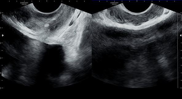
첫 내원 당일 검사한 경직장 전립선 초음파 사진상 사정관 입구의 결석과 탈락된 상피 세포가 쌓여 입구의 직경이 감소하고 있는 초음파 사진입니다.
Transrectal prostate ultrasound image taken on the first visit shows a stone at the opening of the ejaculatory duct
and an accumulation of desquamated epithelial cells, resulting in a reduced diameter of the duct opening.
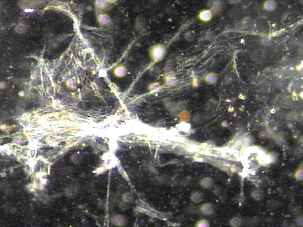
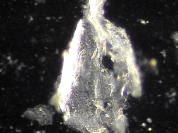
전립선의 표적 치료후 막혀 있던 배출물의 현미경학적 자료입니다.
Microscopic examination of previously obstructed secretions discharged following targeted prostate therapy.
전립선의 표적 치료후 막혀 있던 배출물의 현미경학적 자료입니다.
Microscopic examination of previously obstructed secretions discharged following targeted prostate therapy.
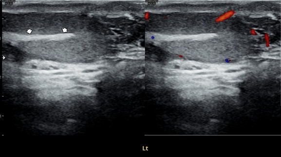
내원 첫날 고환의 초음파 검사상 정관의 순환 장애로 고환 부위에 선상의 고에코가 관찰되는 사진입니다.
During your first visit, an ultrasound of the testicles showed a bright line-like area, which may be caused by poor circulation in the vas deferens.
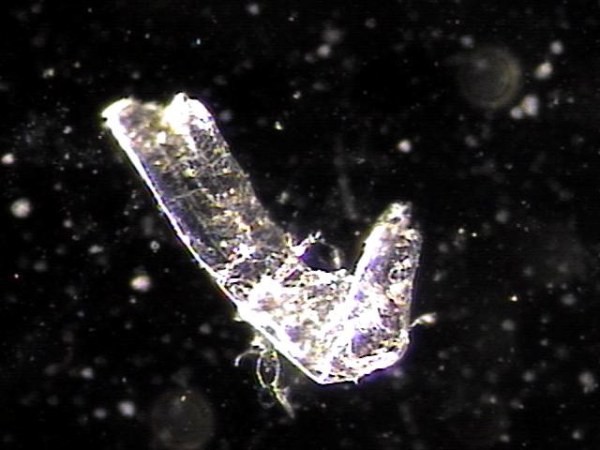
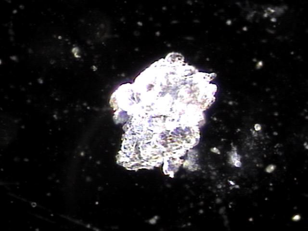
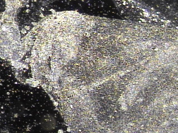
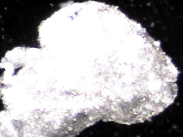
주3회 전립선이 표적 치료후 정관과 사정관 그리고 정낭등에 막혀 있던 탈락된 상피세포 덩어리의 현미경학적 자료 입니다.
This is a microscopic image of exfoliated epithelial cell clusters that were obstructing the vas deferens, ejaculatory ducts, and seminal vesicles, which were released after targeted prostate treatment three times a week.
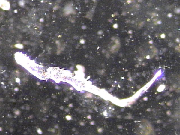
주3회 치료중 정관과 사정관,전립선내 그리고 정낭등에 막혀 있던 섬유소 덩어리가 치료된 현미경학적 사진입니다.
This is a microscopic image showing fibrin clumps that had been blocking the vas deferens, ejaculatory ducts, prostate, and seminal vesicles, which were resolved during the course of targeted treatment three times a week.
주 2~3회 정관의 표적 치료후 정관에 막혀 있던 상피 세포 덩어리가 치료된 현미경학적 자료입니다.
This is a microscopic image taken after targeted treatment of the vas deferens 2 to 3 times per week. It shows that the clumps of shed epithelial cells that had been blocking the vas deferens were successfully cleared.
전립선과 사정관 그리고 정관의 표적 치료후 순환 장애물의 치료된 현미경학적 자료입니다.
This microscopic image shows the treated debris that had been blocking circulation in the prostate, ejaculatory ducts, and vas deferens. After receiving targeted prostate therapy, these blockages were successfully cleared.
전립선과 사정관 그리고 정관의 표적 치료후 순환 장애물의 치료된 현미경학적 자료입니다.
This microscopic image shows the treated debris that had been blocking circulation in the prostate, ejaculatory ducts, and vas deferens. After receiving targeted prostate therapy, these blockages were successfully cleared.
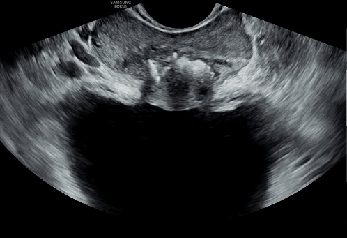
10년 전부터 회음부의 통증과 최근 수년전부터 배뇨장애와 빈뇨가 점점 심해져 다른 비뇨기과의원에서 치료를 했으나 증상의 호전이 없다고 내원하신 당일의 경직장 전립선 초음파 사진 좌우 사정관 입구에 결석이 있고 전립선의 주변구역으로 커져서 전립선을 압박하고 있는 사진입니다.
This transrectal prostate ultrasound image was taken on the patient’s first visit. He had been experiencing perineal pain for the past 10 years, with worsening urinary symptoms and frequency over the recent years. Despite receiving treatment at other urology clinics, his symptoms did not improve. The image shows stones located at the entrances of both ejaculatory ducts, and an enlarged area surrounding the prostate that is pressing against it.
전립선의 표적 치료후 막혀 있던 배출물의 현미경학적 자료입니다.
Microscopic examination of previously obstructed secretions discharged following targeted prostate therapy.
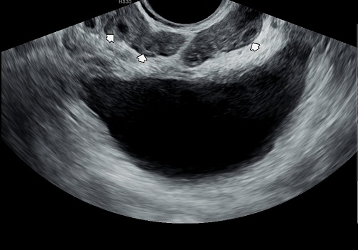
내원 첫날 경직장 전립선 초음파 검사상 정낭의 다발성 낭종이 관찰되는 자료입니다.
On the first day of your visit, the transrectal prostate ultrasound showed multiple cysts in the seminal vesicles.
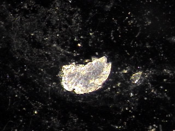
전립선과 정낭, 사정관,전립선관,정관등의 표적 치료후 치료된 상피세포덩어리의 현미경학적 자료입니다.
This microscopic image shows clusters of dead or damaged epithelial cells that were successfully removed through targeted treatment of the prostate, seminal vesicles, ejaculatory ducts, prostate ducts, and vas deferens.
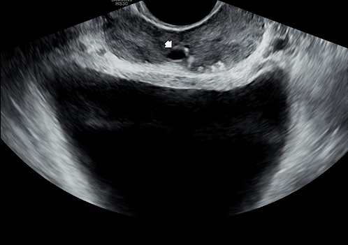
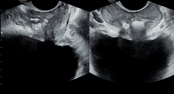
첫 내원당일 경직장 전립선 초음파 사진상 사정관 낭종과 사정관 주위에 쌓여 있는 미세 결석들이 관찰되는 자료입니다.
This ultrasound image, taken during the first visit, shows small cysts and tiny stones gathered around the ejaculatory ducts,
which may be contributing to symptoms.
내원 첫날 경직장 전립선의 측면과 정면 사진상 사정관 입구에 쌓인 결석이 전립선관을 타고 내려가 전립선의 이행 구역를 침범하여 압박과 배뇨장애 빈뇨
그리고 급박뇨등의 증상을 생기게하는 경직장 전립선 초음파 사진입니다.
This transrectal prostate ultrasound image, taken on the first day of the patient's visit, shows stones accumulated at the ejaculatory duct openings. These stones appear to have traveled down through the prostatic ducts, invading the transitional zone of the prostate. This has resulted in pressure on the prostate and symptoms such as urinary difficulties, frequent urination, and urgency.
전립선과 정낭, 사정관,전립선관,정관등의 표적 치료후 치료된 상피세포덩어리의 현미경학적 자료입니다.
This microscopic image shows clusters of dead or damaged epithelial cells that were successfully removed through targeted treatment of the prostate, seminal vesicles, ejaculatory ducts, prostate ducts, and vas deferens.
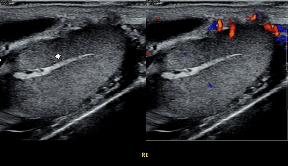
내원 첫날 고환의 초음파 검사상 정관의 순환 장애로 고환 부위에 선상의 고에코가 관찰되는 사진입니다.
During your first visit, an ultrasound of the testicles showed a bright line-like area, which may be caused by poor circulation in the vas deferens.
상기 두 환자분의 병력과 경직장 전립선 초음파 검사들과 전립선과 정낭 그리고 사정관과 정관등의 표적 치료후 결과 오로지 자위 행위등으로 손상 받는 남성 생식기의 자료 입니다.
이러한 비뇨 생식기의 증상과 장기의 손상을 줄이기 위해 건전한 사회 생활과 운동 그리고 가족을 이루도록 노력하여야할 이유입니다.
The medical histories and transrectal prostate ultrasound images of these two patients, along with the results of targeted treatments to the prostate, seminal vesicles, ejaculatory ducts, and vas deferens, all reflect damage to the male reproductive organs caused primarily by excessive self-stimulation."
"To help prevent such urogenital symptoms and organ damage, it is important to pursue a healthy lifestyle, regular exercise, and work toward building meaningful relationships and family life.
댓글목록
등록된 댓글이 없습니다.


