전립선자료실
페이지 정보
본문
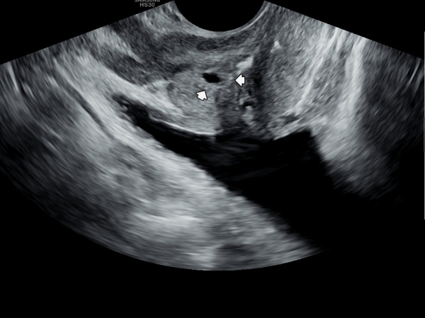
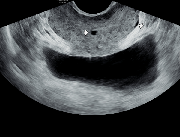
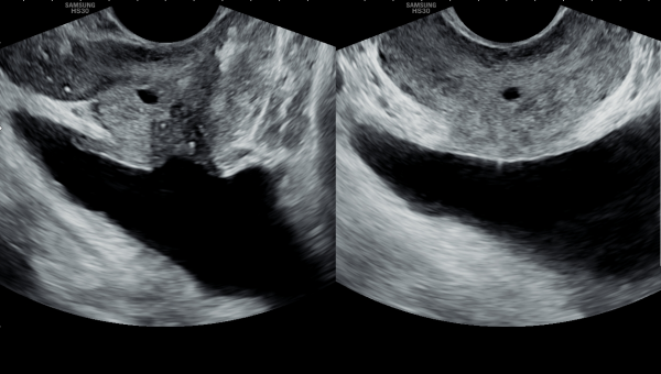
수년전 부터 하복부 통증과 사정시 혈정액을 주소로 여러 비뇨기과와 상급병원에서 치료를 했으나 증상의 호전이 없다고 내원당일 검사한 경직장 전립선 초음파 검사상 사정관 입구에 결석과 사정관의 낭종이 관찰되는 초음파 사진입니다.
The transrectal prostate ultrasound image taken on the day of the patient's first visit shows a stone at the ejaculatory duct opening and a cyst in the ejaculatory duct.
The patient had been experiencing lower abdominal pain and hematospermia during ejaculation for several years, and had received treatment at multiple urology clinics and advanced hospitals without symptom improvement.
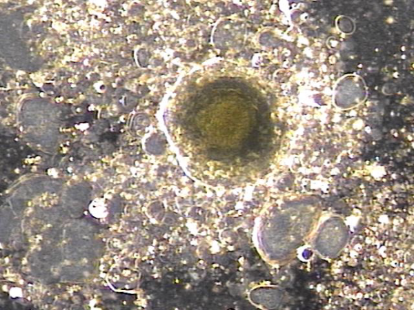
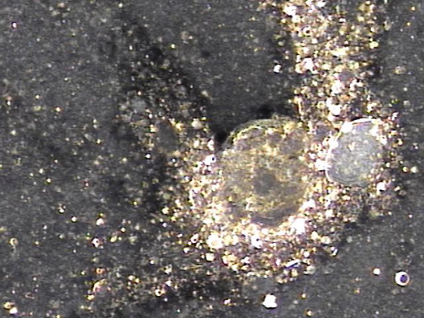
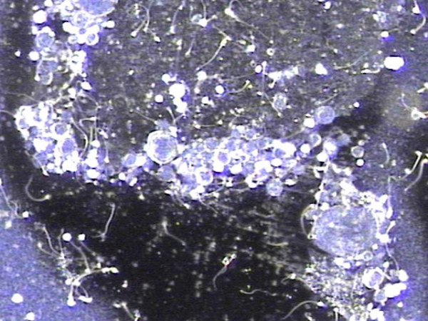
전립선과 정낭과 정관 그리고 사정관 등에서 전립선의 표적 치료후 배양 검사를 하기위해 배출된 결석과 혈정액의 현미경 학적 사진입니다.
Microscopic image of stones and hematospermia discharged from the prostate, seminal vesicles, vas deferens, and ejaculatory ducts following targeted prostate therapy, collected for culture testing.
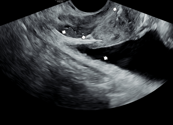
내원당일 경직장 전립선 초음파 검사상 정낭의 낭종이 커져 있고 정낭내 결석이 관찰되는 측면 경직장 초음파 사진입니다.
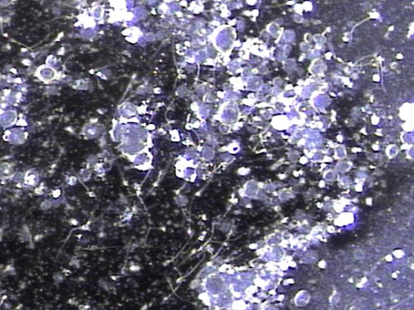
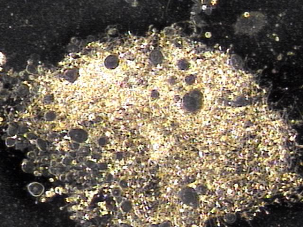
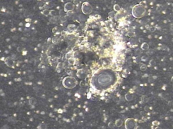
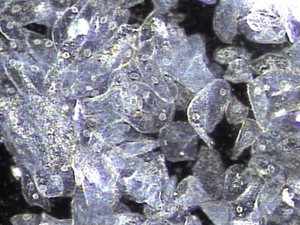
주2 ~3회 서울가정의학과에서 전립선과 사정관, 정관 그리고 정낭등의 표적 치료후 수명을 다하고 탈락되어 정관과 사정관 그리고 전립선관 등에 막혀 있던 탈락된 상피세포와 치료된 혈정액의 현미경학적 사진입니다.
This is a microscopic image of detached epithelial cell clusters and treated hematospermia that were discharged following targeted treatment of the prostate, ejaculatory duct, vas deferens, and seminal vesicles, performed 2 to 3 times per week at Seoul Family Medicine Clinic. These epithelial cells had reached the end of their life cycle and were obstructing the vas deferens, ejaculatory duct, and prostatic ducts prior to treatment.
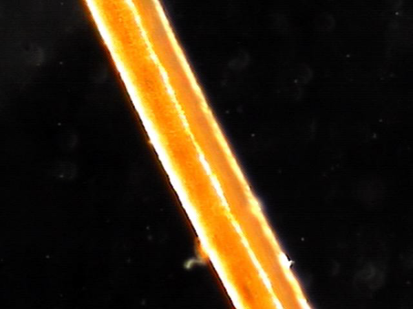
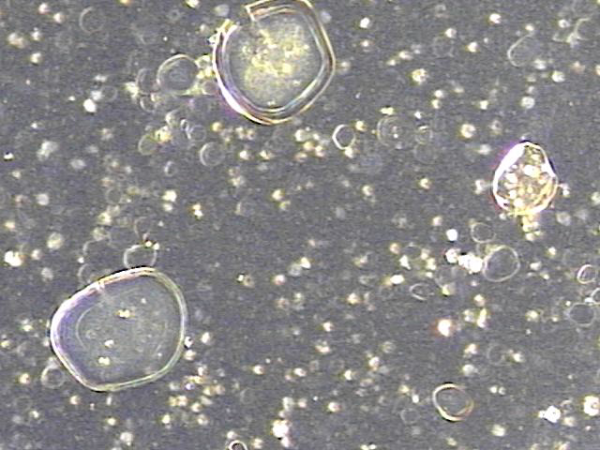
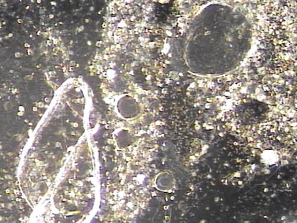
전립선과 사정관과 정낭 그리고 정관등이 표적치료를 한다음 배출된 정낭의 결석과 혈정액의 현미경 학적 검사 자료상 머리카락의 굵기와 비교하여 치료를 하므로 열심히 치료하여 건강하게 삶을 삽시다.
This is a microscopic examination of seminal vesicle stones and hematospermia discharged after targeted treatment of the prostate, ejaculatory ducts, seminal vesicles, and vas deferens. By comparing their size to the thickness of a human hair, we can see the importance of precise treatment. Let’s continue treatment diligently and live a healthy life.
첫 내원 당일 정면 경직장 전립선 초음파 사진상 좌우 사정관 입구에 결설이 관찰되고 전립선의 결절이 관찰되는 초음파 사진입니다.
This is a frontal transrectal prostate ultrasound image taken on the patient's first visit, showing calculi (stones) at the openings of both ejaculatory ducts and nodules within the prostate.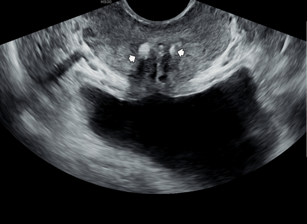
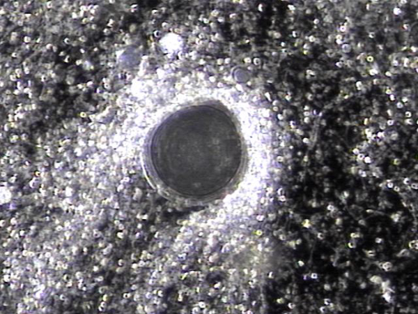
내원 첫날 전립선의 표적 치료후 막혀 있던 사정관과 정낭등의 결석이 배출되고 혈정액등이 치료되고 있는 현미경학적 자료입니다.
This is a microscopic image taken after the first targeted prostate treatment on the day of the initial visit, showing the discharge of calculi that had been blocking the ejaculatory ducts and seminal vesicles, as well as the treatment of hemospermia and related findings.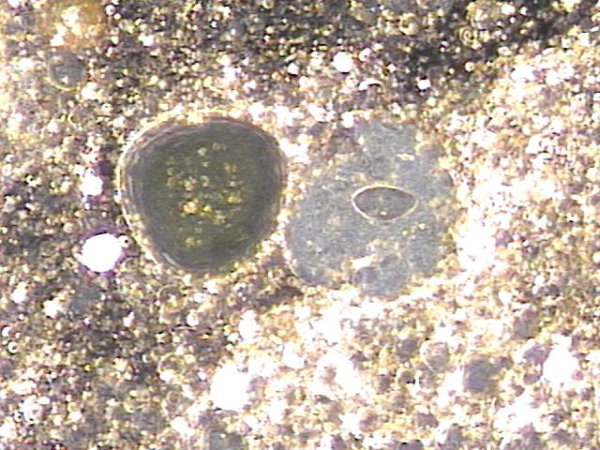
내원 첫날 정면 경직장 전립선 초음파 사진상 사정관 입구가 순환장애로 사정관의 낭종과 부분적 전립선 낭종들이 관찰되는 초음파 사진입니다.
내원 첫날 전립선의 표적 치료후 막혀 있던 사정관과 정낭등의 결석이 배출되고 혈정액등이 치료되고 있는 현미경학적 자료입니다.
This is a microscopic image taken after the first targeted prostate treatment on the day of the initial visit, showing the discharge of calculi that had been blocking the ejaculatory ducts and seminal vesicles, as well as the treatment of hemospermia and related findings.
일주일동안 주 3회 전립선,사정관,정관 그리고 정낭의 표적 치료후 배출된 정낭의 결석의 현미경학적 검사 자료 입니다.
This is a microscopic examination of seminal vesicle calculi and hematospermia discharged following one week of targeted therapy—administered three times per week—on the prostate, ejaculatory ducts, vas deferens, and seminal vesicles.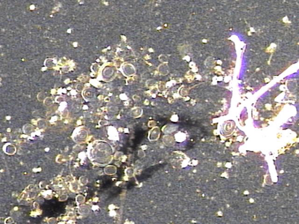
내원 첫날 경직장 전립선 초음파 검사상 양측 사정관에 막혀 있던 탈락된 상피 세포와 결석들이 쌓여서 항문 주위에 채워져 나중 급박뇨와 빈뇨와 요실금등의 원인이 되는 사진입니다.
This is a transrectal prostate ultrasound image taken on the first day of the visit, showing accumulated desquamated epithelial cells and calculi obstructing both ejaculatory ducts. These blockages extend around the anal region and are likely contributing to symptoms such as urinary urgency, frequency, and incontinence.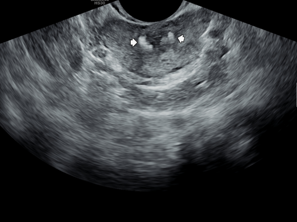
내원 첫날 전립선의 표적 치료후 막혀 있던 사정관과 정낭등의 결석이 배출되고 혈정액등이 치료되고 있는 현미경학적 자료입니다.
This is a microscopic image taken after the first targeted prostate treatment on the day of the initial visit, showing the discharge of calculi that had been blocking the ejaculatory ducts and seminal vesicles, as well as the treatment of hemospermia and related findings.
일주일동안 주 3회 전립선,사정관,정관 그리고 정낭의 표적 치료후 배출된 정낭의 결석의 현미경학적 검사 자료 입니다.
This is a microscopic examination of seminal vesicle calculi and hematospermia discharged following one week of targeted therapy—administered three times per week—on the prostate, ejaculatory ducts, vas deferens, and seminal vesicles.
수년전 부터 하복부 통증과 사정시 혈정액을 주소로 여러 비뇨기과와 상급병원에서 치료를 했으나 증상의 호전이 없다고 내원당일 검사한 경직장 전립선 초음파 검사상 사정관 입구에 결석과 사정관의 낭종이 관찰되는 초음파 사진입니다.
The transrectal prostate ultrasound image taken on the day of the patient's first visit shows a stone at the ejaculatory duct opening and a cyst in the ejaculatory duct.
The patient had been experiencing lower abdominal pain and hematospermia during ejaculation for several years, and had received treatment at multiple urology clinics and advanced hospitals without symptom improvement.
주2 ~3회 서울가정의학과에서 전립선과 사정관, 정관 그리고 정낭등의 표적 치료후 수명을 다하고 탈락되어 정관과 사정관 그리고 전립선관 등에 막혀 있던 탈락된 상피세포와 치료된 혈정액의 현미경학적 사진입니다.
This is a microscopic image of detached epithelial cell clusters and treated hematospermia that were discharged following targeted treatment of the prostate, ejaculatory duct, vas deferens, and seminal vesicles, performed 2 to 3 times per week at Seoul Family Medicine Clinic. These epithelial cells had reached the end of their life cycle and were obstructing the vas deferens, ejaculatory duct, and prostatic ducts prior to treatment.
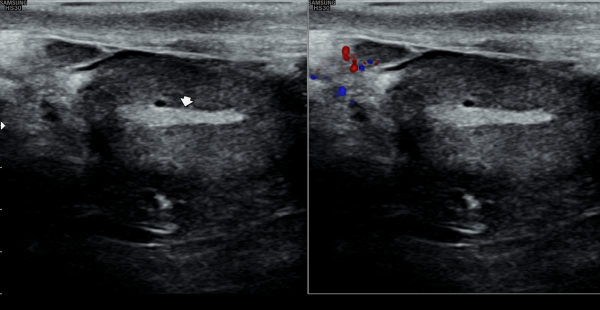
수년동안 사정관과 정관등의 순환장애로 내원 당일 고환의 초음파 검사상 고환의 섬유화가 관찰되는 초음파 사진입니다.
This is an ultrasound image of the testes taken on the day of the visit, showing testicular fibrosis likely caused by years of circulatory disturbances in the ejaculatory ducts and vas deferens.
주2 ~3회 서울가정의학과에서 전립선과 사정관, 정관 그리고 정낭등의 표적 치료후 수명을 다하고 탈락되어 정관과 사정관 그리고 전립선관 등에 막혀 있던 탈락된 상피세포와 치료된 혈정액의 현미경학적 사진입니다.
This is a microscopic image of detached epithelial cell clusters and treated hematospermia that were discharged following targeted treatment of the prostate, ejaculatory duct, vas deferens, and seminal vesicles, performed 2 to 3 times per week at Seoul Family Medicine Clinic. These epithelial cells had reached the end of their life cycle and were obstructing the vas deferens, ejaculatory duct, and prostatic ducts prior to treatment.
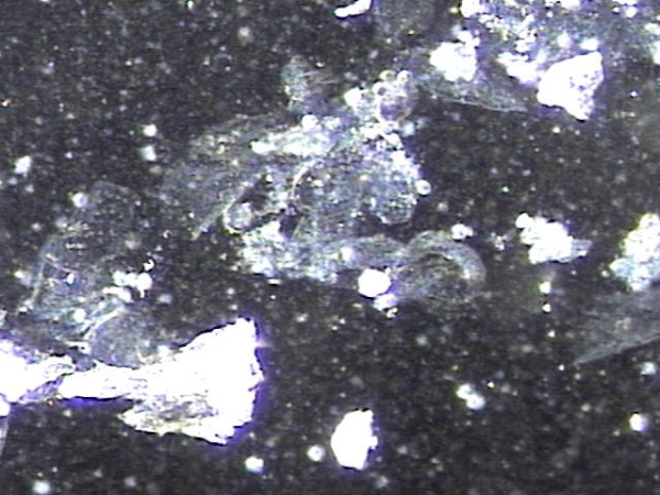
주 3회 전립선과 정낭 그리고 사정관등의 표적치료후 치료된 혈정액과 정낭 결석등의 현미경학적 자료입니다.
This is a microscopic examination image of treated hematospermia and seminal vesicle calculi, obtained after targeted therapy of the prostate,
seminal vesicles, and ejaculatory ducts administered three times per week.
주 3회 전립선과 정낭 그리고 사정관등의 표적치료후 치료된 혈정액과 정낭 결석등의 현미경학적 자료입니다.
This is a microscopic examination image of treated hematospermia and seminal vesicle calculi, obtained after targeted therapy of the prostate,
seminal vesicles, and ejaculatory ducts administered three times per week.
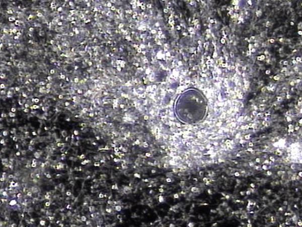
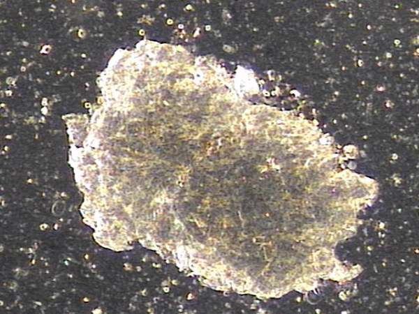
주3회 전립선과 사정관의 표적치료후 치료된 상피세포덩어리의 현미경학적 검사 자료입니다.
This is a microscopic examination image of a cluster of epithelial cells that were treated and discharged following targeted therapy of the prostate and ejaculatory ducts, performed three times a week.
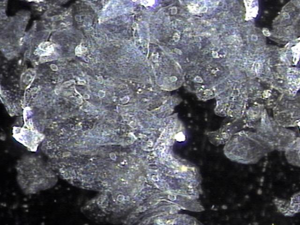
주3회 전립선과 사정관의 표적치료후 치료된 상피세포와 결석들의 현미경학적 검사 자료입니다.
This is a microscopic examination image of a cluster of epithelial cells with microcalculi that were treated and discharged following targeted therapy of the prostate and ejaculatory ducts, performed three times a week.
주 3회 전립선의 표적 치료시 상피 세포 덩어리가 배출된 현미경학적 자료 입니다.
Microscopic findings showing the expulsion of epithelial cells clusters during targeted prostate therapy administered three times per week.
주 3회 전립선의 표적 치료시 오래된 상피 세포 덩어리가 배출된 현미경학적 자료 입니다.
Microscopic findings showing the expulsion of old epithelial cell clusters during targeted prostate therapy administered three times per week.
댓글목록
등록된 댓글이 없습니다.


