전립선자료실
페이지 정보
본문
수년전부터 회음부 통증과 배뇨 장애로 내원 당일 검사한 경직장 전립선 초음파 검사상 사정관 입구의 미세 결석과
전립선의 낭종이 관찰되는 초음파 사진입니다.
A transrectal prostate ultrasound image taken on the day of the visit shows microcalcifications at the opening of the ejaculatory duct and cysts within the prostate, in a patient who had been experiencing perineal pain and voiding difficulties for several years.
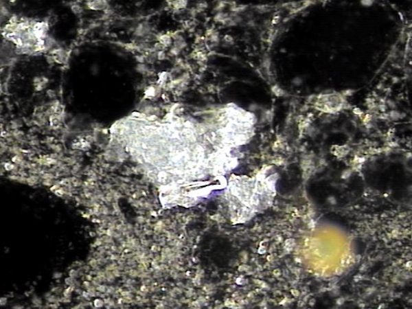
내원당일 전립선의 표적 치료후 배양과 전립선액의 PCR 검사를 위해 채취하고 검사한 현미경학적 확대 사진입니다.
A high-magnification microscopic image taken on the day of the visit, following targeted prostate therapy,
showing the prostatic fluid sample collected for culture and PCR testing.
5년전부터 폐암으로 투병중 전립선 치료를 받지 못하던중 최근 하복부 통증이 심해져 내원 당일 추적 경직장 전립선 초음파 추적 검사상
전립선 낭종들이 커지고 사정관 입구의 미세 결석이 관찰되는 사진입니다.
A follow-up transrectal prostate ultrasound image taken on the day of the visit shows enlargement of prostatic cysts and microcalcifications at the opening of the ejaculatory duct in a patient who had been unable to receive prostate treatment due to lung cancer for the past five years and recently developed worsening lower abdominal pain.
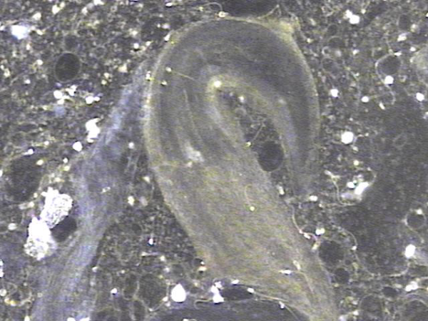
주 2회 전립선의 표적 치료후 배출된 상피세포의 현미경학적 사진입니다.
A microscopic image of epithelial cells expelled after twice-weekly targeted prostate therapy.
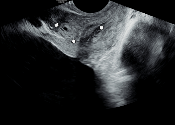
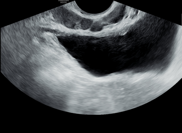
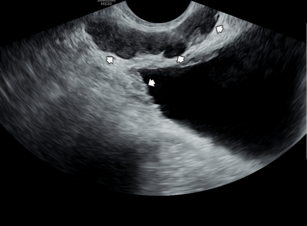
5년전 내원 당일 정낭의 초음파 검사상 좌측 정낭 낭종이 의심되는 사진입니다.
An ultrasound image of the seminal vesicles taken on the day of the visit five years ago, showing a suspected cyst in the left seminal vesicle.
주 2회 전립선의 표적 치료후 배출된 상피세포의 현미경학적 사진입니다.
A microscopic image of epithelial cells expelled after twice-weekly targeted prostate therapy.

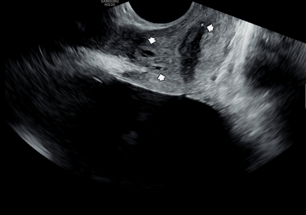
5년후 내원 당일 정낭의 경직장 전립선 초음파 검사상 정낭의 낭종들이 커지고 있는 사진입니다.
A transrectal prostate ultrasound image of the seminal vesicles taken on the day of the visit five years later, showing enlargement of seminal vesicle cysts.
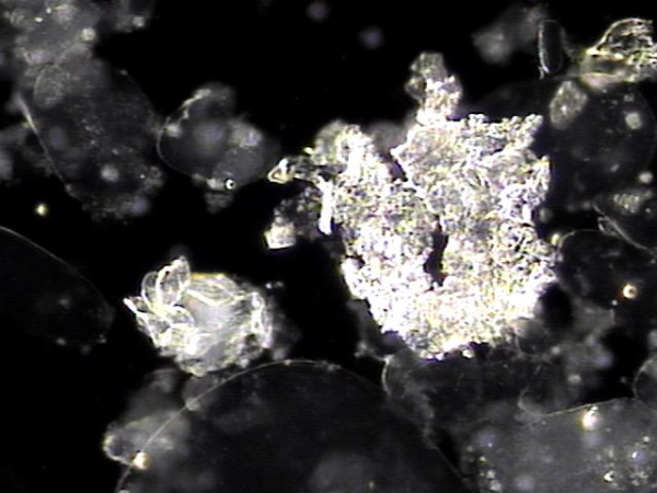
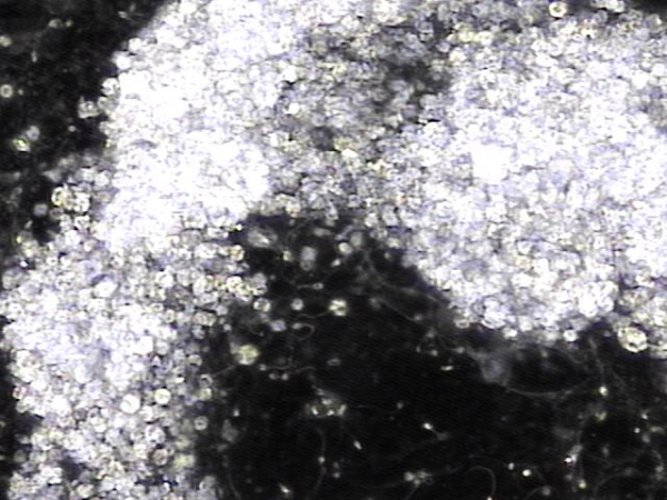
주 2회 전립선의 표적 치료후 배출된 염증덩어리의 현미경학적 사진입니다.
A microscopic image of an inflammatory mass expelled after twice-weekly targeted prostate therapy.
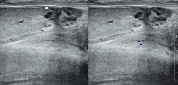
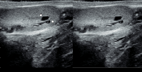
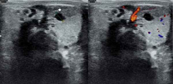
5년전 내원 당일 고환의 초음파 검사상 고환 낭종이 관찰되는 초음파 사진입니다.
A testicular ultrasound image taken on the day of the visit five years ago, showing a testicular cyst.
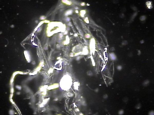
주2회 전립선의 표적 치료후 배출된 섬유들의 현미경학적 자료입니다.
Microscopic findings of fibrous tissue expelled after twice-weekly targeted prostate therapy.
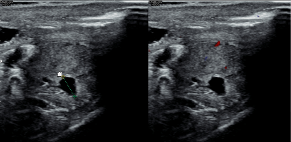
5년 지나 정관의 순환장애로 생긴 고환 낭종이 커지고 있는 초음파 사진입나디.
An ultrasound image showing an enlarging testicular cyst caused by impaired circulation in the vas deferens, five years later.
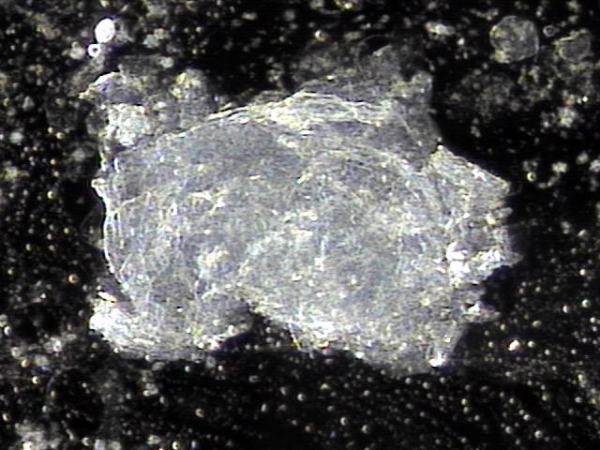
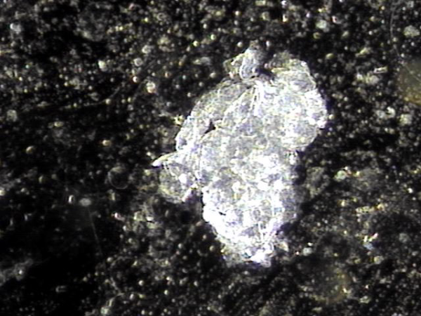
주 2회 전립선, 정관,정낭,그리고 사정관등의 표적 치료후 치료된 상피세포 덩어리의 현미경학적 사진입니다.
This is a microscopic image of epithelial cell clusters treated after targeted therapy twice a week on the prostate, vas deferens, seminal vesicles, and ejaculatory ducts.
5년전 내원 당일 고환의 초음파 검사상 고환 낭종이 관찰되는 사진입니다.
This is an ultrasound image showing a testicular cyst observed on the day of the visit 5 years ago.
주 2회 전립선, 정관,정낭,그리고 사정관등의 표적 치료후 치료된 상피세포 덩어리의 현미경학적 사진입니다.
This is a microscopic image of epithelial cell clusters treated after targeted therapy twice a week on the prostate, vas deferens, seminal vesicles, and ejaculatory ducts.
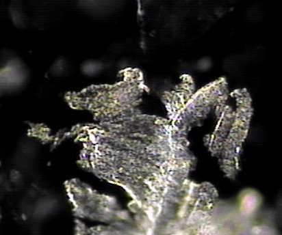
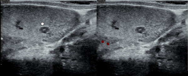
5년후 추적 고환의 초음파 검사상 정관의 순환 장애로 고환 낭종이 커지고 있는 초음파 사진입니다.
This is a follow-up ultrasound image of the testis 5 years later, showing enlargement of the testicular cyst due to circulatory disturbance of the vas deferens.
주 2회 전립선, 정관,정낭,그리고 사정관등의 표적 치료후 치료된 상피세포 덩어리의 현미경학적 사진입니다.
This is a microscopic image of epithelial cell clusters treated after targeted therapy twice a week on the prostate, vas deferens, seminal vesicles, and ejaculatory ducts.
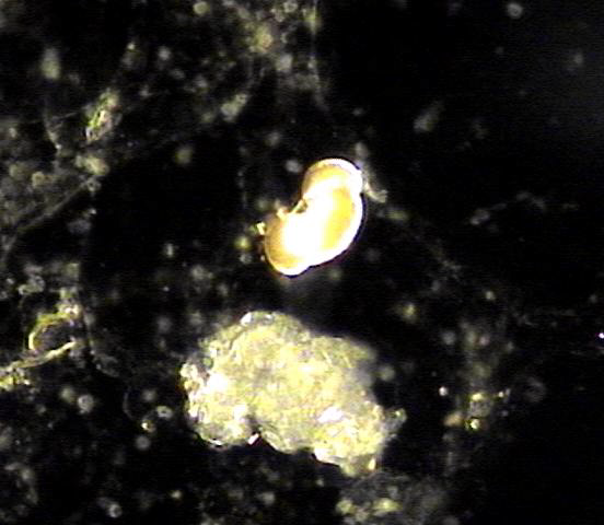
주 2회 전립선, 정관,정낭,그리고 사정관등의 표적 치료후 치료된 상피세포 덩어리와 정관에 막혀 있던 결절의 현미경학적 사진입니다.
This is a microscopic image of treated epithelial cell clusters and nodules that were obstructing the vas deferens, following targeted therapy twice a week on the prostate, vas deferens, seminal vesicles, and ejaculatory ducts.
댓글목록
등록된 댓글이 없습니다.


