전립선자료실
페이지 정보
본문
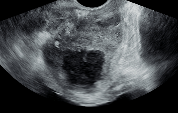
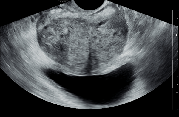
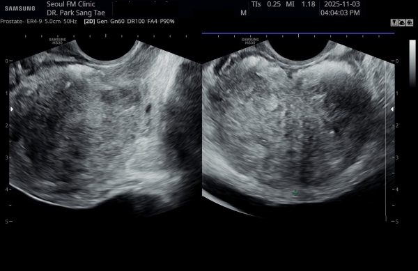
17개월동안 상급 의료 기관에서 전립선 치료에 대한 투약을 했으나 증상의 호전이 없이 배뇨장애와 배뇨시 통증으로
내원 당일 경직장 전립선 초음파 사진상 전립선내 거대 농양이 관찰되는 자료입니다.
"A transrectal prostate ultrasound image taken on the day of the visit, showing a large prostatic abscess in a patient
who had been receiving prostate treatment with medication at a higher-tier medical institution for 17 months without symptomatic improvement,
experiencing voiding dysfunction and pain during urination."
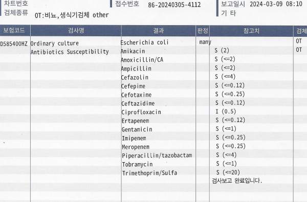
내원 당일 전립선의 표적 치료후 배출된 전립선액의 배양 검사와 항생제 민감도 검사 결과
"Culture test and antibiotic sensitivity test results of prostatic fluid expelled after targeted prostate therapy on the day of the visit."
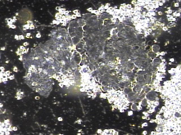
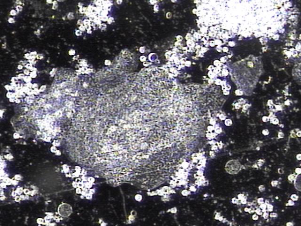
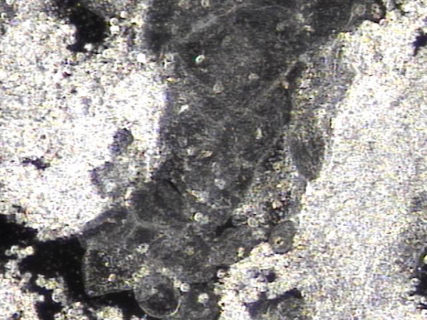
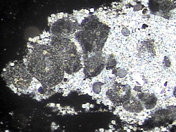
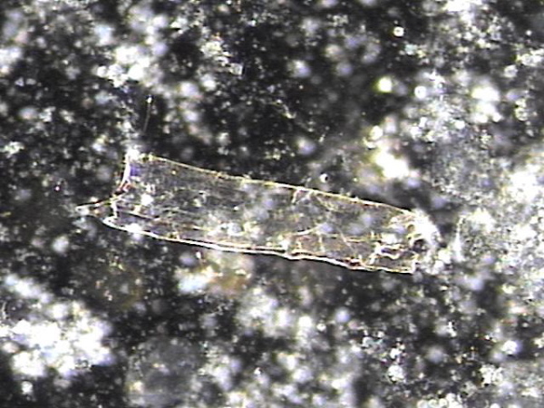
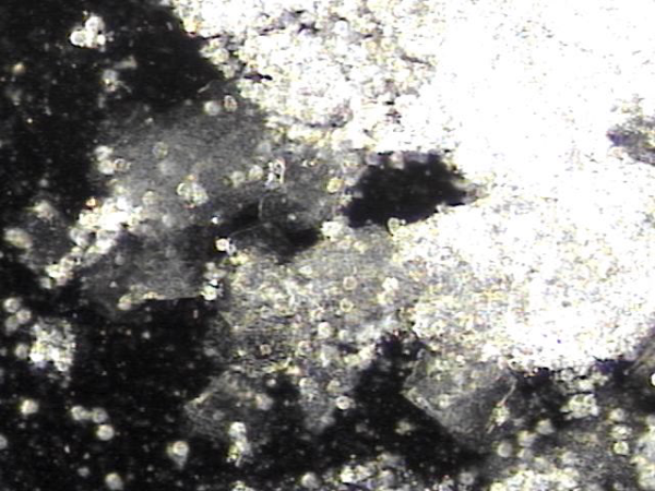
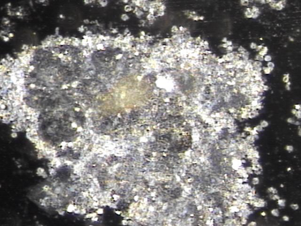
주2회 전립선의 표적 치료후 탈락된 상피세포 덩어리와 염증세포들입니다.
"Desquamated epithelial cell clusters and inflammatory cells after twice-weekly targeted prostate therapy."
주2회 전립선의 표적 치료후 탈락된 상피세포 덩어리와 염증세포들입니다.
"Desquamated epithelial cell clusters and inflammatory cells after twice-weekly targeted prostate therapy."
주2회 전립선의 표적 치료후 탈락된 상피세포 덩어리와 염증세포들입니다.
"Desquamated epithelial cell clusters and inflammatory cells after twice-weekly targeted prostate therapy."
주2회 전립선의 표적 치료후 탈락된 상피세포 덩어리와 염증세포들입니다.
"Desquamated epithelial cell clusters and inflammatory cells after twice-weekly targeted prostate therapy."
주2회 전립선의 표적 치료후 탈락된 상피세포 덩어리와 염증세포들입니다.
"Desquamated epithelial cell clusters and inflammatory cells after twice-weekly targeted prostate therapy."
주2회 전립선의 표적 치료후 탈락된 상피세포 덩어리와 염증세포 그리고 정자들이 모여 있는 정낭액의 사진입니다 .
"Desquamated epithelial cell clusters and inflammatory cells after twice-weekly targeted prostate therapy."
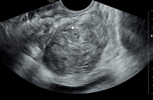
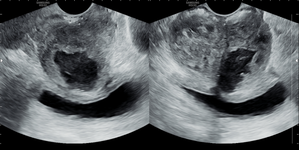
주 2회 전립선의 표적 치료후 전립선의 농양이 없어진 경직장 전립선의 추적 검사 사진.
"A follow-up transrectal prostate ultrasound image showing the resolution of a prostatic abscess
after twice-weekly targeted prostate therapy."
주 2회 전립선과 사정관,정낭 그리고 정관등의 표적 치료후 전립선내 커다란 낭종이 치료된 추적 경직장 전립선 초음파 사진입니다.
This follow-up transrectal prostate ultrasound image shows that, after twice-weekly targeted treatment of the prostate, ejaculatory ducts, seminal vesicles, and vas deferens, a large cyst inside the prostate has improved.
주 2회 전립선의 표적 치료후 전립선의 농양이 없어지고 전립의 크기가 줄고 결석이 감소하고 있는 경직장 전립선의 추적 검사 사진.
"A follow-up transrectal prostate ultrasound image showing the resolution of the prostatic abscess, a reduction in prostate size,
and a decrease in calculi after twice-weekly targeted prostate therapy."
주 1회 전립선의 표적 치료후 전립선과 사정관,정낭 그리고 정관등에서 치료된 상피세포 덩어리와 염증 세포들의 현미경 학적 자료입니다.
This is a microscopic image taken after once- or twice-weekly targeted treatment of the prostate, ejaculatory ducts, seminal vesicles, and vas deferens. It shows clumps of shed epithelial cells and inflammatory cells that have been successfully treated.
세균의 배양 검사상 전립선내 농양에 있는 세균이 치료된 배양 검사 결과지.
"A bacterial culture test result showing the eradication of bacteria in the prostatic abscess
after twice-weekly targeted prostate therapy.
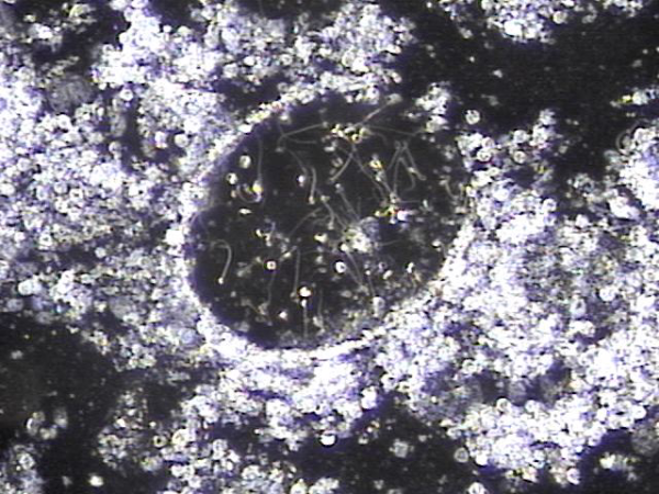
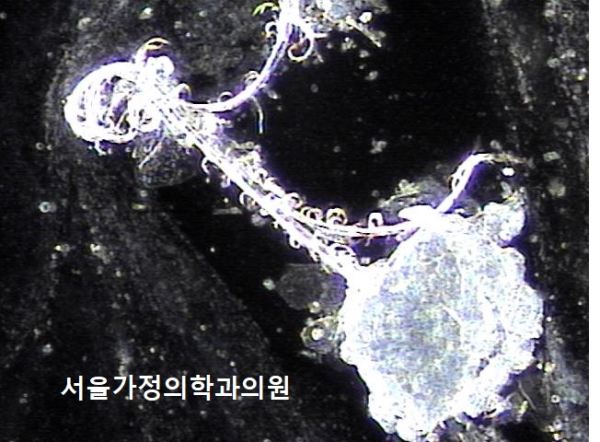
주 1회 전립선의 표적 치료중이라도 하루 약 300만 마리의 정자가 고환에서 생산되어 사정관이 막혀 정자의 순환 장애로 정낭에 모여 혈정액 혹은 정낭 결석과 하복부 통증 그리고 만성 골반통 증후군등의
원인의 치료된 현미경학적 자료입니다.
Even though the testes naturally produce about 3 million sperm every day, blockages in the ejaculatory ducts can sometimes prevent sperm from flowing properly. This can cause semen to build up in the seminal vesicles, leading to blood in the semen, stones, lower abdominal pain, or chronic pelvic pain. Thanks to the targeted treatments, these blockages are being cleared, and this microscopic image shows the tissue after successful treatment.
주 1회 치료중 정낭에서 생산되는 염증 즉 프로스타그란딘 R로 생긴 염증 덩어리와 탈락된 거짓 중층 원주 상피세포의 치료된 현미경 학적 자료입니다.
This microscopic image shows the treated tissue from the seminal vesicles. It includes inflammatory clumps caused by prostaglandin-related inflammation, along with shed epithelial cells that had been blocking the ducts. These findings show that the treatment is helping to clear away these causes and improve circulation.
주 1회 치료중 정낭에서 생산되는 염증 즉 프로스타그란딘 R로 생긴 염증 덩어리와 탈락된 거짓 중층 원주 상피세포의 치료된 현미경 학적 자료입니다.
This microscopic image shows the resolution of inflammatory clusters, caused by prostaglandin R produced in the seminal vesicles, along with shed pseudostratified columnar epithelial cells, following once- or twice-weekly treatment.
주 1 치료중 막혀 있던 사정관내 탈락된 상피 세포와 프로스타그란딘에 의한 염증 세포 덩어리가 치료된 현미경학적 자료 입니다.
This microscopic image shows tissue and inflammatory cell clusters that were blocking the ejaculatory duct. These materials, including shed epithelial cells and inflammation caused by prostaglandins, were cleared after twice-weekly targeted treatments.
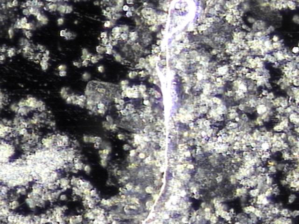
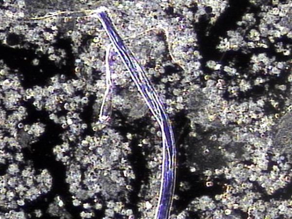
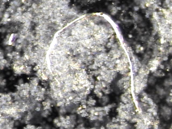
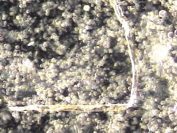
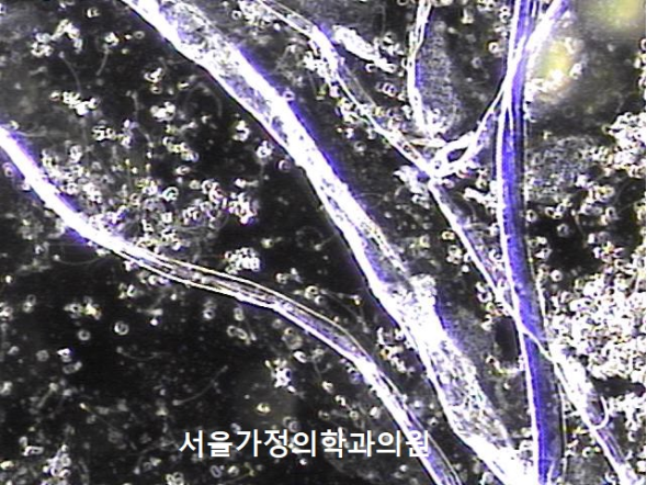
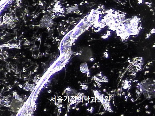
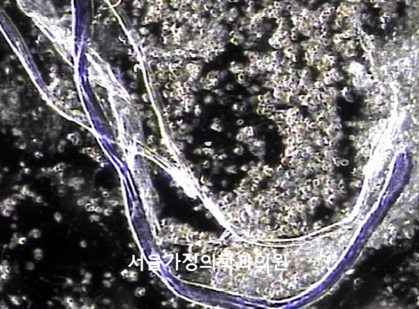
전립선과 정낭, 정관과 사정관 그리고 전립선관등의 표적 치료후 치료된 사정관과 정관 그리고 전립선관내 막혀있던 상피 세포 찌꺼기와 단백질 그리고 노페물의 현미경학적 자료 입니다.
This microscopic image shows materials that were blocking the ejaculatory duct, vas deferens, and prostatic ducts—such as shed epithelial cells, protein debris, and waste substances. These materials were released and cleared following targeted treatments of the prostate, seminal vesicles, and related ducts, helping to restore better flow and function.
전립선과 정낭 그리고 정관등의 순환키위해 탈락된 상피세포와 단백질과 노페물이 전립선관과 사정관 그리고 정관등으로 배출되고 있는 현미경학적 자료입니다.
This microscopic image shows material that was cleared from the vas deferens and related ducts after targeted treatment — including aggregated epithelial cell debris, proteinaceous matter, and inflammatory components. These substances appear to have been blocking the normal flow and may have contributed to symptoms. The fact that they were expelled confirms that the ducts are responding to treatment and opening up.
사정관내 정관내 그리고 전립선관내 막혀 있던 탈락된 상피 세포들과 단백질과 노폐물등의 현미경학적 자료입니다.
This microscopic image shows material that was cleared from the vas deferens and related ducts after targeted treatment — including aggregated epithelial cell debris, proteinaceous matter, and inflammatory components. These substances appear to have been blocking the normal flow and may have contributed to symptoms. The fact that they were expelled confirms that the ducts are responding to treatment and opening up.
전립선내 선상피들이 탈락되어 전립선관내로 배출되고 있는 현미경학적 자료입니다.
This microscopic image shows the shedding of glandular epithelial cells from the prostate into the duct. This process is part of the natural healing and cleansing response, helping to remove damaged or inflamed cells and allowing the prostate ducts to open and function more smoothly.
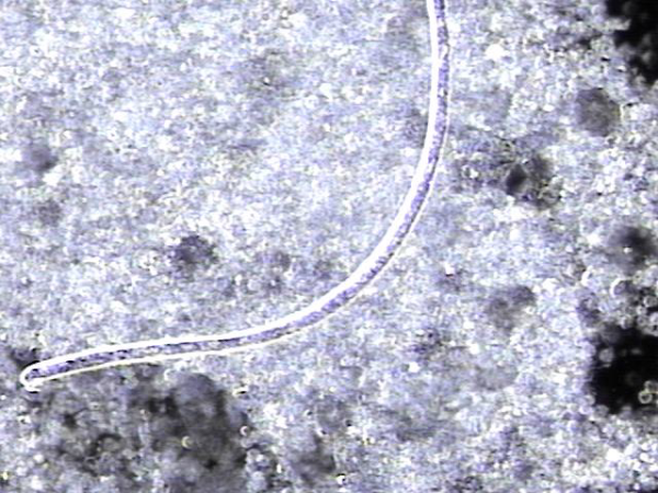
주1회 전립선과 정낭 그리고 사정관과 정관등의 표적 치료후 탈락된 상피세포와 농세포들의 치료된 현미경학적 자료입니다.
These are microscopic findings showing treated and detached epithelial cells and pus cells observed after one to two sessions per week of targeted therapy to the prostate, seminal vesicles, ejaculatory ducts, and vas deferens.
댓글목록
등록된 댓글이 없습니다.


