전립선 자료실
페이지 정보
본문
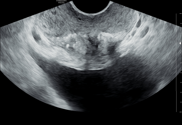
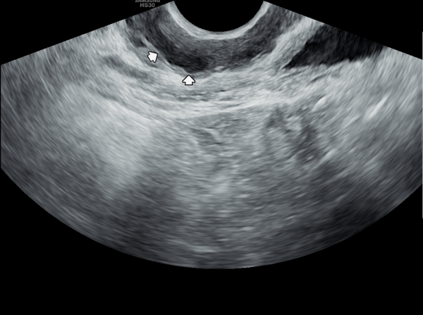
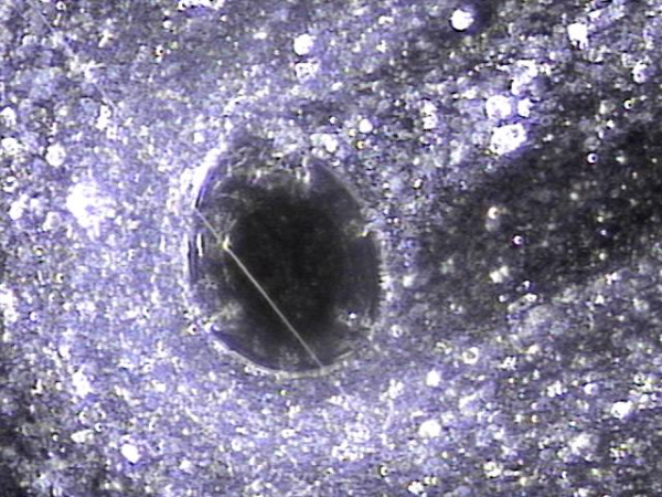
수년전부터 하복부 통증과 배뇨장애와 빈뇨와 급밥뇨가 심해 개인 비뇨기과와 상급의료기관에서 치료를 했으나 증상의 호전이 없고 최근 두달전 증상이 심해져 내원 당일 경직장 초음파 검사상 좌우 사정관 입구의 결석이 심하고 오래전부터 막혀 전립선의 이행구역과 주변 구역까지 결석이 침습된 전립선 사진입니다.
For several years, the patient had been experiencing lower abdominal pain, urinary dysfunction, frequent urination, and urgency. Despite receiving treatment at private urology clinics and tertiary medical centers, there was no improvement in symptoms. On the day of the visit, a transrectal ultrasound examination revealed severe calcification at the openings of both ejaculatory ducts. The ducts had been chronically obstructed, allowing the calcifications to invade the transition zone and surrounding areas of the prostate.
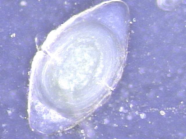
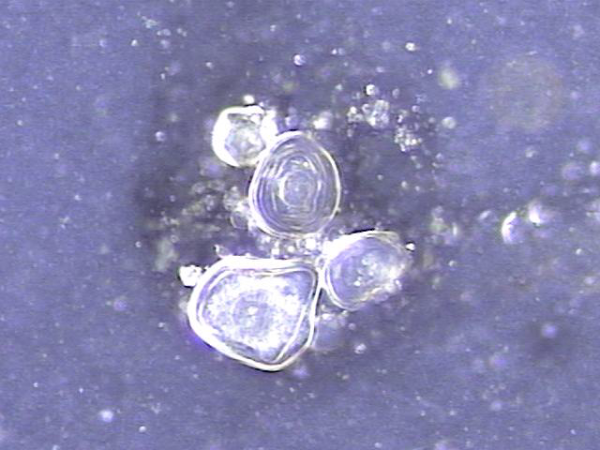
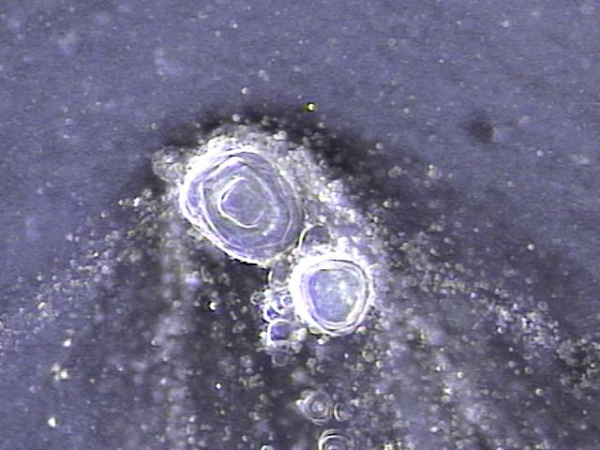
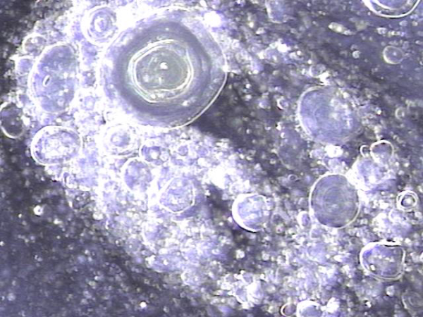
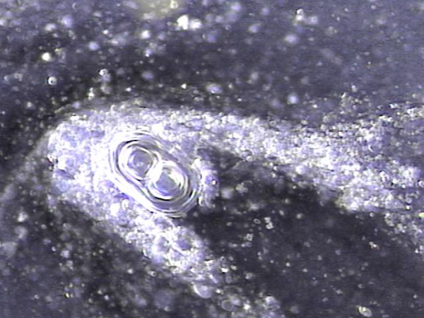
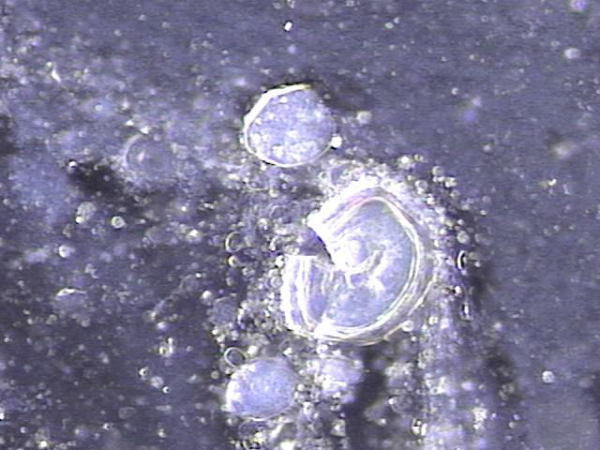
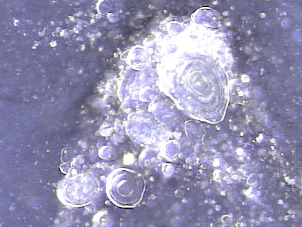
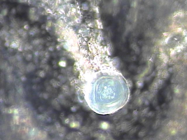
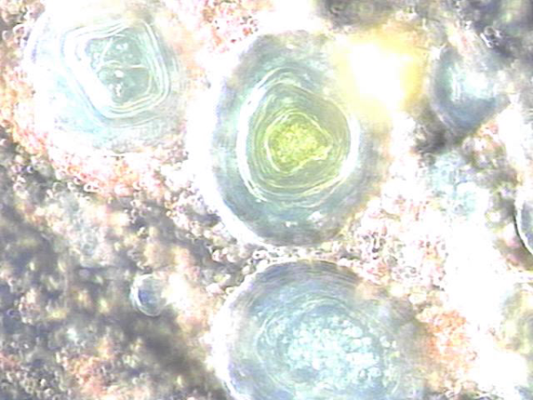
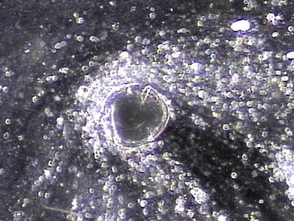
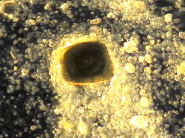
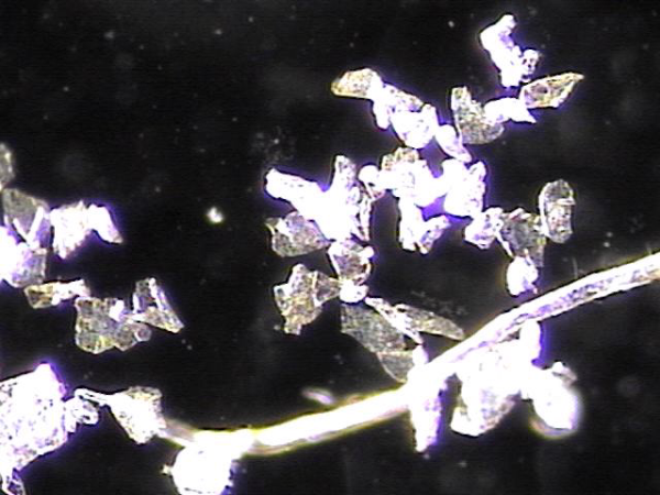
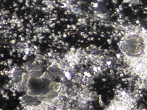
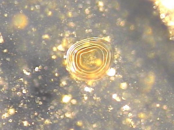
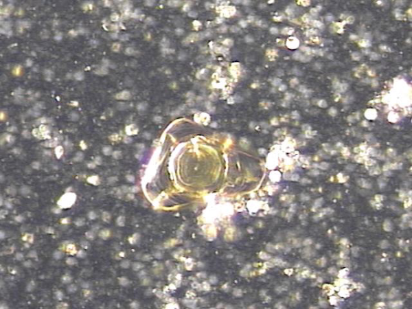
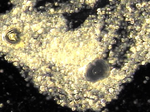
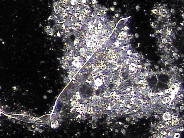
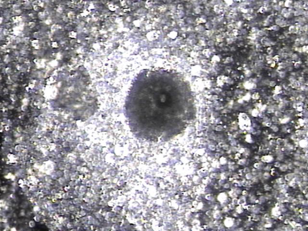
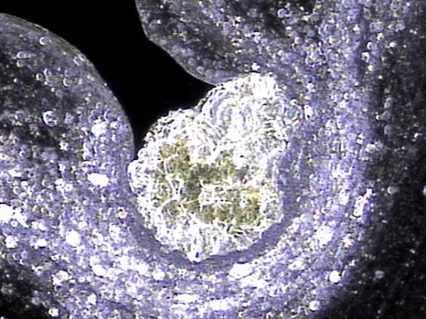
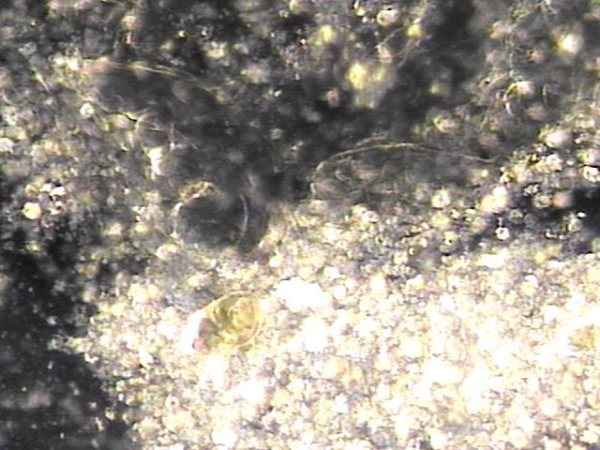
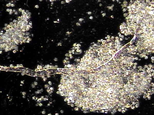
내원 당일 전립선의 표적 치료후 배출된 전립선액의 배양과 PCR 검사를 한후 현미경학적 검사상 치료된 사정관 결석과 전립선의 결석 자료입니다.
On the day of the visit, following targeted prostate treatment, prostatic fluid was discharged and analyzed through culture and PCR testing. Microscopic examination revealed treated ejaculatory duct stones and prostatic calculi.
수년전부터 하복부 통증과 배뇨장애와 빈뇨와 급밥뇨가 심해 개인 비뇨기과와 상급의료기관에서 치료를 했으나 증상의 호전이 없고 최근 두달전 증상이 심해져 내원 당일 경직장 초음파 검사상 좌우 사정관 입구의 결석이 심하고 오래전부터 막혀 전립선의 이행구역과 주변 구역까지 결석이 침습된 전립선 사진입니다.
For several years, the patient had been experiencing lower abdominal pain, urinary dysfunction, frequent urination, and urgency. Despite receiving treatment at private urology clinics and tertiary medical centers, there was no improvement in symptoms. On the day of the visit, a transrectal ultrasound examination revealed severe calcification at the openings of both ejaculatory ducts. The ducts had been chronically obstructed, allowing the calcifications to invade the transition zone and surrounding areas of the prostate.
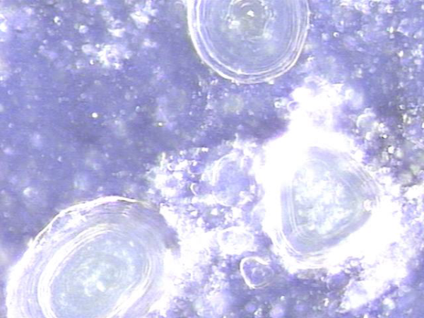
내원 당일 전립선의 표적 치료후 배출된 전립선액의 배양과 PCR 검사를 한후 현미경학적 검사상 치료된 사정관 결석과 전립선의 결석 자료입니다.
On the day of the visit, following targeted prostate treatment, prostatic fluid was discharged and analyzed through culture and PCR testing. Microscopic examination revealed treated ejaculatory duct stones and prostatic calculi.
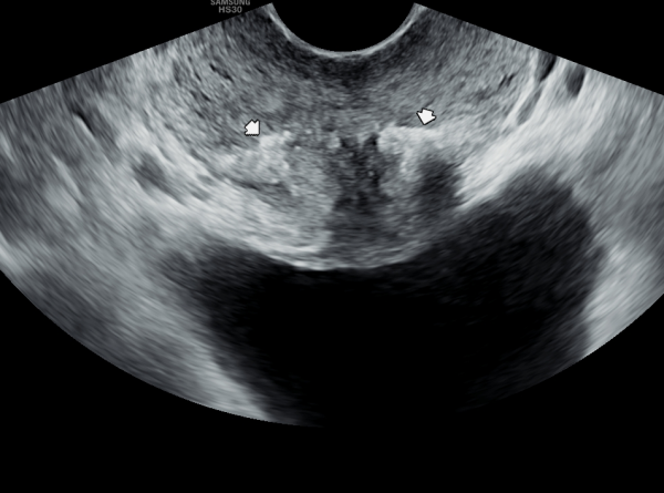
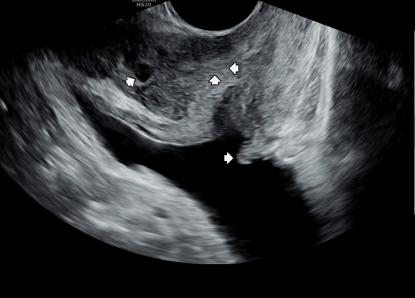
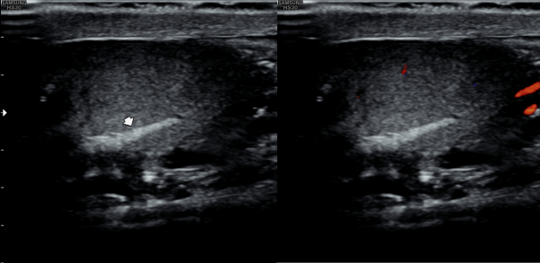
첫 내원 당일 측면 경직장 전립선 초음파 검사상 사정관 입구의 결석과 폐쇄로 사정관 낭종이 관찰되고 사정관에 탈락된 상피세포가 쌓여 사정관이 막히고 있으며 요도 협착등 배뇨 장애로 방관내 조직의 이상 증식이 관찰되는 초음파 사진 입니다.
On the first day of the visit, a lateral transrectal prostate ultrasound revealed stones and obstruction at the entrance of the ejaculatory ducts, leading to ejaculatory duct cysts. Detached epithelial cell debris was observed accumulating and blocking the ducts. In addition, abnormal tissue proliferation within the bladder was noted, likely due to urethral stricture and associated voiding dysfunction.
내원 당일 전립선의 표적 치료후 배출된 전립선액의 배양과 PCR 검사를 한후 현미경학적 검사상 치료된 사정관 결석과 전립선의 결석 자료입니다.
On the day of the visit, following targeted prostate treatment, prostatic fluid was discharged and analyzed through culture and PCR testing. Microscopic examination revealed treated ejaculatory duct stones and prostatic calculi.
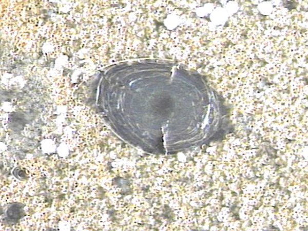
전립선과 사정관 그리고 사정관입구의 결석 치료중 사정관의 좁은 입구와 전립선관의 막혀 있는 입구로 커다란 결석이 배출시 압력으로 좁아져 있거나
막혀 있던 섬유화된 입구의 손상이 예상되는 현미경학적 사진입니다.
This is a microscopic image taken during the treatment of stones in the prostate, ejaculatory ducts, and the ejaculatory duct orifices.
It shows that large stones being expelled through the narrowed or obstructed fibrotic openings of the ejaculatory and prostatic ducts likely caused mechanical damage due to pressure during expulsion.
내원 당일 정낭의 경직장 전립선 초음파 검사상 사정관입구의 섬유화와 탈락된 상피 세포 그리고 결석에 의한 폐쇄로 정관을 통한 정자의 순환 장애로 커진 정낭과 혈정액등을 일으키는 초음파 사진입니다.
This is a transrectal prostate ultrasound image of the seminal vesicles taken on the day of the patient's visit. The image shows fibrotic changes
at the ejaculatory duct openings, along with obstructive epithelial debris and calculi, leading to impaired sperm flow through the vas deferens,
resulting in enlarged seminal vesicles and hematospermia.
내원 당일 전립선의 표적 치료후 치료된 전립선 결석과 혈정액의 현미경 학적 자료입니다.
This is a microscopic image of treated prostatic calculi and hematospermia collected after targeted prostate therapy on the day of the patient’s initial visit.
내원 당일 전립선의 표적 치료후 배출된 전립선액의 배양과 PCR 검사를 한후 현미경학적 검사상 치료된 사정관 결석과 전립선의 결석 자료입니다.
On the day of the visit, following targeted prostate treatment, prostatic fluid was discharged and analyzed through culture and PCR testing. Microscopic examination revealed treated ejaculatory duct stones and prostatic calculi.
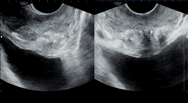
내원 당일 측면 경직장 전립선 초음파 사진과 정면 경직장 전립선 초음파 사진입니다.
These are the sagittal and frontal transrectal ultrasound images of the prostate taken on the day of the initial visit.
내원 당일 전립선의 표적 치료후 치료된 전립선 결석과 혈정액의 현미경 학적 자료입니다.
This is a microscopic image of treated prostatic calculi and hematospermia collected after targeted prostate therapy on the day of the patient’s initial visit.
내원 당일 전립선의 표적 치료후 배출된 전립선액의 배양과 PCR 검사를 한후 현미경학적 검사상 치료된 사정관 결석과 전립선의 결석 자료입니다.
On the day of the visit, following targeted prostate treatment, prostatic fluid was discharged and analyzed through culture and PCR testing. Microscopic examination revealed treated ejaculatory duct stones and prostatic calculi.
내원 당일 고환의 초음파 사진상 사정관의 결석과 섬유화로 정관의 순환 장애를 일으켜 좌우 고환의 섬유화가 생기고 있는 고환 초음파 사진입니다.
This is a scrotal ultrasound image taken on the day of the initial visit, showing fibrosis in both testes caused by obstructed circulation in the vas deferens due to calcifications and fibrosis of the ejaculatory ducts.
내원 당일 전립선의 표적 치료후 치료된 전립선 결석과 혈정액의 현미경 학적 자료입니다.
This is a microscopic image of treated prostatic calculi and hematospermia collected after targeted prostate therapy on the day of the patient’s initial visit.
전립선과 사정관 그리고 사정관입구의 결석 치료중 사정관의 좁은 입구와 전립선관의 막혀 있는 입구로 커다란 결석이 배출시 압력으로 좁아져 있거나
막혀 있던 섬유화된 입구의 손상이 예상되는 현미경학적 사진입니다.
This is a microscopic image taken during the treatment of stones in the prostate, ejaculatory ducts, and the ejaculatory duct orifices.
It shows that large stones being expelled through the narrowed or obstructed fibrotic openings of the ejaculatory and prostatic ducts likely caused mechanical damage due to pressure during expulsion.
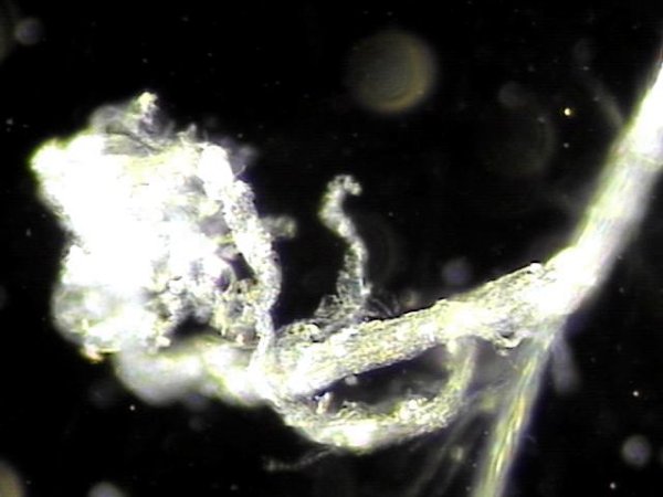
탈락된 상피세포로 좁아진 사정관과 전립선관 그리고 사정관 입구의 결석들이 배출시 손상된 조직 치료되고 전립선의 표적치료후 치료된 전립선과 사정관 결석과 염증세포 덩어리의 현미경학적 자료입니다.
This is a microscopic image showing treated prostatic and ejaculatory duct stones, along with clusters of inflammatory cells, after targeted therapy of the prostate. It illustrates the healing of tissues damaged during the expulsion of dislodged epithelial cells, which had narrowed the ejaculatory ducts, prostatic ducts, and ejaculatory duct openings.
주 3회 전립선의 표적 치료후 치료된 상피 세포들의 현미경학적 사진 힙니다.
This is a microscopic image of the epithelial cells treated following targeted prostate therapy performed three times a week.
주3회 전립선의 표적 치료후 사정관과 전립선관 그리고 정관등에 막혀 있던 오래된 상피세포 덩어리의 현미경학적 자료입니다.
Microscopic findings of old epithelial cell clusters that had been obstructing the ejaculatory ducts, prostatic ducts,
and vas deferens, observed after targeted prostate therapy administered three times per week.
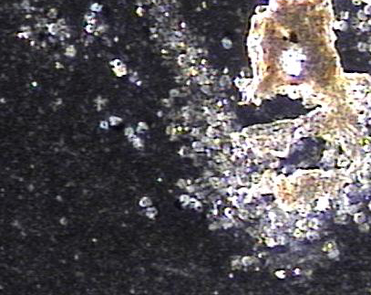
탈락된 상피세포로 좁아진 사정관과 전립선관 그리고 사정관 입구의 결석들이 배출시 손상된 조직 치료되고 전립선의 표적치료후 치료된 전립선과 사정관 결석과 염증세포 덩어리의 현미경학적 자료입니다.
This is a microscopic image showing treated prostatic and ejaculatory duct stones, along with clusters of inflammatory cells, after targeted therapy of the prostate. It illustrates the healing of tissues damaged during the expulsion of dislodged epithelial cells, which had narrowed the ejaculatory ducts, prostatic ducts, and ejaculatory duct openings.
전립선의 주 3회 표적치료후 전립선관과 사정관, 그리고 정관등에 막혀 있던 섬유소 덩어리의 치료된 현미경학적 자료입니다.
This is a microscopic image of treated fibrin clumps that were blocking the prostatic ducts, ejaculatory ducts, and vas deferens, following targeted therapy on the prostate three times a week.
탈락된 상피세포로 좁아진 사정관과 전립선관 그리고 사정관 입구의 결석들이 배출시 손상된 조직들이 전립선의 표적 치료후 전립선관내 그리고 사정관과 그 입구를 막고있는 결석과 프로스타그란딘으로 생긴 염증 세포 덩어리들이 치료된 현미경학적 자료입니다.
This microscopic image shows tissue that had been damaged when stones and shed epithelial cells blocked the ejaculatory ducts, prostatic ducts, and their openings. After targeted prostate treatment, the stones and inflammatory cell clusters caused by prostaglandins were successfully treated and cleared.
탈락된 상피세포로 좁아진 사정관과 전립선관 그리고 사정관 입구의 결석들이 배출시 손상된 조직들이 전립선의 표적 치료후 전립선관내 그리고 사정관과 그 입구를 막고있는 결석과 프로스타그란딘으로 생긴 염증 세포 덩어리들이 치료된 현미경학적 자료입니다.
Here we can see under the microscope how blockages made of stones, old cells, and inflammation inside the prostate and ejaculatory ducts have been cleared after targeted treatment. This shows the healing process and improvement in circulation.
전립선관과 사정관 그리고 정낭등에 막혀 있던 프로스타그란딘으로 생긴 염증덩어리와 체위 요법과 표적 치료로 치료된 결석들의 현미경학적 자료입니다.
This microscopic image shows small inflammatory clusters and stones that were blocking the prostate ducts and surrounding areas. With targeted therapy and positional exercises, these blockages have been successfully treated and improved.
주 2회 사정관입구와 사정관, 전립선관,정낭 그리고 정관등의 표적 치료후 치료된 현미경학적 자료입니다.
주 2회 전립선관과 사정관, 정낭 그리고 정관등의 표적치료후 치료된 현미경학적 자료입니다.
Here is microscopic evidence obtained after twice-weekly targeted therapy of the prostatic ducts, ejaculatory ducts, seminal vesicles, and vas deferens.
댓글목록
등록된 댓글이 없습니다.


