전립선자료실
페이지 정보
본문
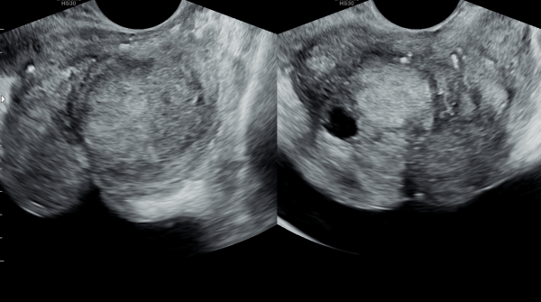
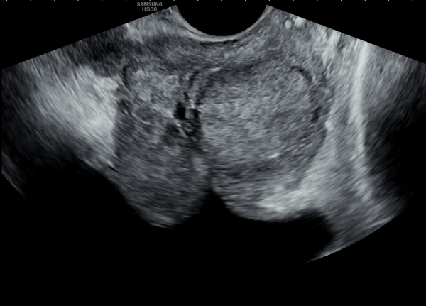
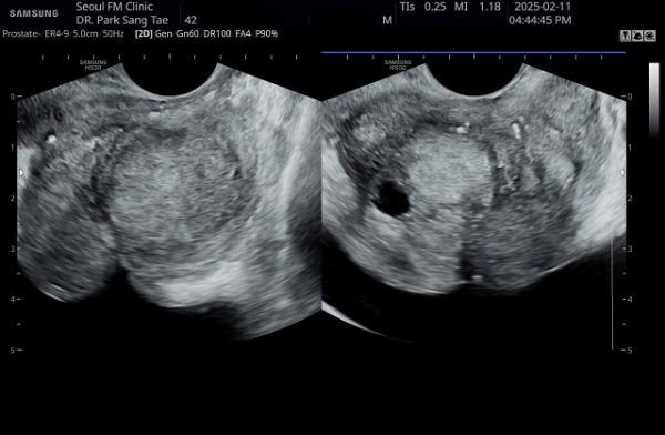
10년전부터 배뇨장애와 빈뇨로 비뇨기과에서 약을 복용중 2년전부터 급박뇨와 요실금이 심해져 투약을 했으나 증상의 호전이 없다고 내원당일 검사한 경직장 전립선 초음파 검사상 전립선 비대와 전립선의 낭종과
사정관주위의 결석이 관찰되는 경직장 전립선 초음파 사진입니다.
This is a transrectal prostate ultrasound image taken on the day of the visit. The patient had been taking medication for urinary difficulties and frequent urination for over 10 years at a urology clinic. However, since two years ago, symptoms of urgency and urinary incontinence worsened despite continued medication. The ultrasound shows prostate enlargement, cysts within the prostate, and calcifications around the ejaculatory ducts.
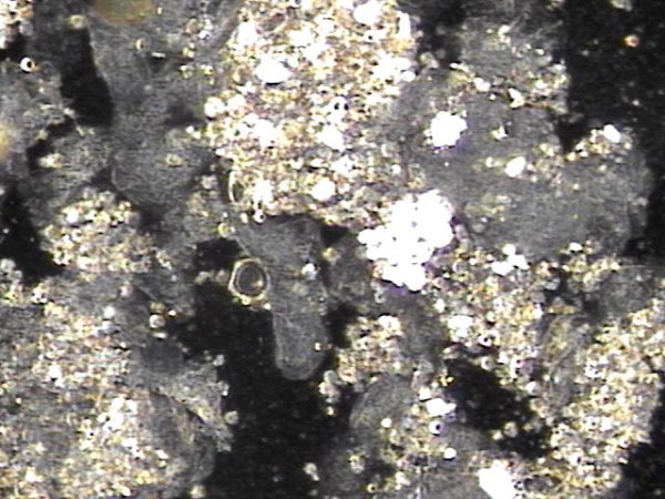
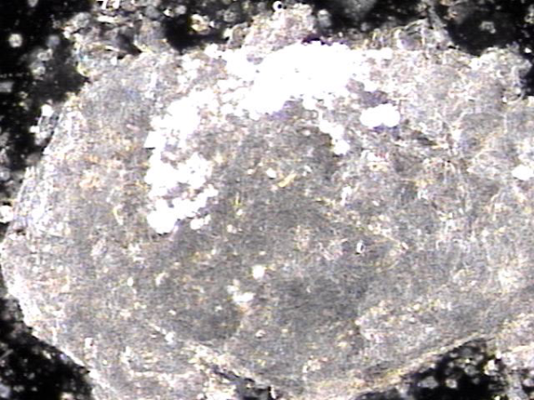
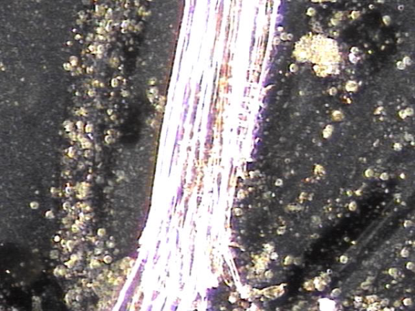
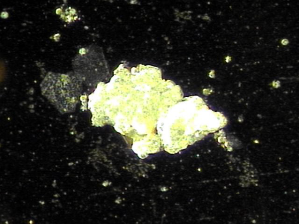
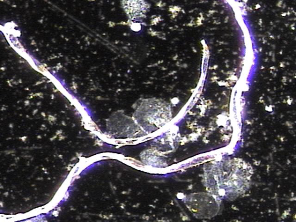
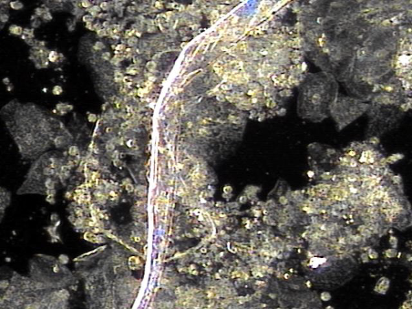
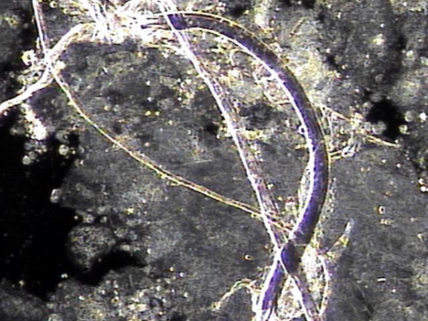
주 2회 전립선의 표적 치료중 전립선액과 정낭액 그리고 전립선 결석과 정관등에 침범하여 퍼지고 있는 균의 배양과 항생제 민감도 검사를 하기위해 표적 치료후 배출된 전립선액의 현미경 검사 자료입니다.
탈락된 상피세포 덩어리와 혈정액과 전립선 결석과 전립선암의 전암병변 의심되는 자료입니다.
This is a microscopic examination of prostatic fluid discharged during biweekly targeted prostate therapy. The sample was obtained to conduct bacterial culture and antibiotic sensitivity testing for organisms invading and spreading through the prostatic fluid, seminal vesicle fluid, prostate calculi, and vas deferens. The findings include clusters of desquamated epithelial cells, hematospermia, prostatic calculi, and features suspicious for precancerous lesions of prostate cancer.
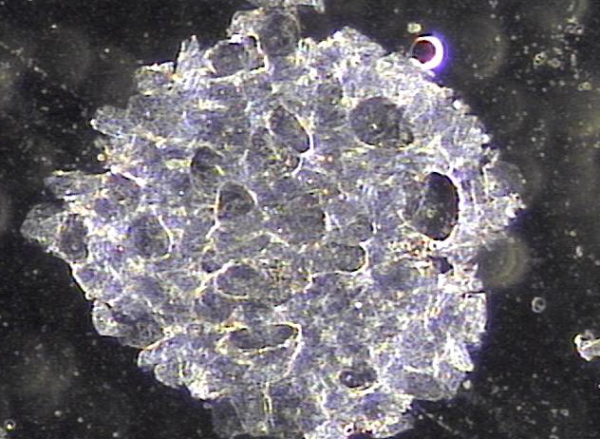
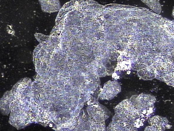
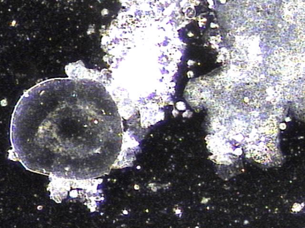
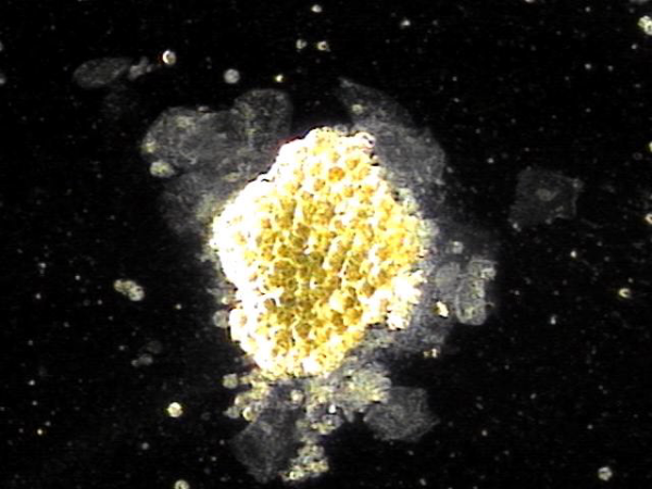
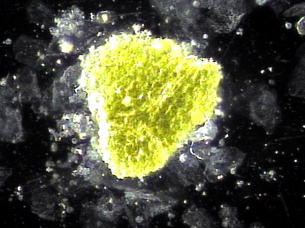
주2회 전립선의 표적 치료후 치료된 상피 세포 덩어리의 현미경학적 자료입니다.
"This is the microscopic data of the epithelial cell clusters treated after targeted prostate therapy twice a week."
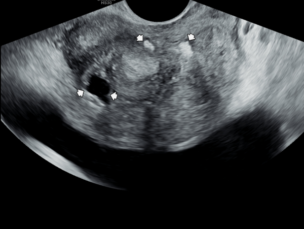
내원 당일 전립선의 정면 초음파 사진상 좌우 사정관 입구의 결석과 전립선의 이행구역에 비대해진 결절 그리고 우측 전립선 결절내 전립선 낭종이 관찰되며
방광쪽으로 커져 배뇨장애와 급박뇨가 심해 지고 있는 경직장 전립선 초음파 사진입니다.
This is a transrectal prostate ultrasound image taken on the day of the patient's first visit, showing stones at the openings of both ejaculatory ducts, an enlarged nodule in the transitional zone of the prostate, and a prostatic cyst within the right prostatic nodule. The prostate is enlarged toward the bladder, contributing to worsening voiding dysfunction and urgency.
주2회 전립선의 표적 치료후 치료된 상피 세포 덩어리의 현미경학적 자료입니다.
"This is the microscopic data of the epithelial cell clusters treated after targeted prostate therapy twice a week."
주2회 전립선의 표적 치료후 치료된 상피 세포 덩어리의 현미경학적 자료입니다.
"This is the microscopic data of the epithelial cell clusters treated after targeted prostate therapy twice a week."
내원 당일 전립선의 정면 초음파 사진상 좌우 사정관 입구의 결석과 전립선의 이행구역에 비대해진 결절 그리고 우측 전립선 결절내 전립선 낭종이 관찰되며
방광쪽으로 커져 배뇨장애와 급박뇨가 심해 지고 있는 경직장 전립선 초음파 사진입니다.
This is a transrectal prostate ultrasound image taken on the day of the patient's first visit, showing stones at the openings of both ejaculatory ducts, an enlarged nodule in the transitional zone of the prostate, and a prostatic cyst within the right prostatic nodule. The prostate is enlarged toward the bladder, contributing to worsening voiding dysfunction and urgency.
주2회 전립선의 표적 치료후 치료된 상피 세포 덩어리의 현미경학적 자료입니다.
"This is the microscopic data of the epithelial cell clusters treated after targeted prostate therapy twice a week."
주2회 전립선의 표적 치료후 치료된 상피 세포 덩어리의 현미경학적 자료입니다.
"This is the microscopic data of the epithelial cell clusters treated after targeted prostate therapy twice a week."
주2회 전립선의 표적 치료후 치료된 상피 세포 덩어리의 현미경학적 자료입니다.
"This is the microscopic data of the epithelial cell clusters treated after targeted prostate therapy twice a week."
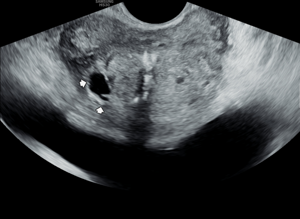
내원 당일 전립선의 측면 초음파 사진상 좌우 사정관 입구의 결석과 전립선의 이행구역에 비대해진 결절 그리고 우측 전립선 결절내 전립선 낭종이 관찰되며
방광쪽으로 커져 배뇨 장애와 급박뇨가 심해 지고 있는 경직장 전립선 초음파 사진입니다.
This is a transrectal prostate ultrasound image taken on the day of the patient's first visit, showing stones at the openings of both ejaculatory ducts, an enlarged nodule in the transitional zone of the prostate, and a prostatic cyst within the right prostatic nodule. The prostate is enlarged toward the bladder, contributing to worsening voiding dysfunction and urgency.
주2회 전립선의 표적 치료후 치료된 상피 세포 덩어리의 현미경학적 자료입니다.
"This is the microscopic data of the epithelial cell clusters treated after targeted prostate therapy twice a week."
주2회 전립선의 표적 치료후 치료된 상피 세포 덩어리의 현미경학적 자료입니다.
"This is the microscopic data of the epithelial cell clusters treated after targeted prostate therapy twice a week."
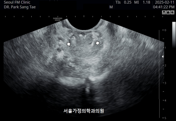
수십년간 빈뇨가 심해 비뇨기과에서 약만 투약하였으나 최근 2년전부터 급박뇨가 심해 내원 당일 검사한 경직장 전립선 초음파 검사상 항문 주위에 쌓인 탈락된
상피세포와 단백질등의 화학적 작용을 결석들이 생겨 항문 올림근등의 기능이 감소하여 급박뇨가 심해지고 있는 초음파 사진입니다.
This ultrasound image, taken during a transrectal prostate examination, shows small calcified deposits and accumulated epithelial cell debris around the anal area. These materials may have developed over time due to chronic inflammation and reduced circulation.
The deposits can affect the pelvic muscles, including the levator ani, leading to reduced muscle control and symptoms such as frequent urination and urgency. This finding helps explain the cause of the patient’s persistent urinary symptoms.
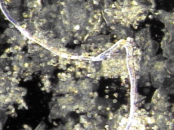
전립선과 정낭,사정관과 정관등의 표적치료후 치료된 탈락되어 막혀 있던 상피세포 덩어리의 현미경 학적 자료입니다.
This is a microscopic image of detached epithelial cell clusters that were blocking the prostate, seminal vesicles, ejaculatory ducts, and vas deferens, now treated through targeted therapy.
전립선과 정낭,사정관과 정관등의 표적치료후 치료된 탈락되어 막혀 있던 상피세포 덩어리의 현미경 학적 자료입니다.
This is a microscopic image of detached epithelial cell clusters that were blocking the prostate, seminal vesicles, ejaculatory ducts, and vas deferens, now treated through targeted therapy.
내원 당일 검사한 고환의 초음파 검사상 고환의 섬유화가 진행된 초음파 사진입니다.
This ultrasound image shows changes in the left testicle. The tissue pattern suggests that fibrosis, or scarring of the testicular tissue, has developed.
Fibrosis often happens after repeated inflammation, past infections, or reduced blood circulation in the area. It is usually a long-term change in the tissue.
At this stage, the finding itself is not dangerous, but it may be related to symptoms such as discomfort, changes in testicular size, or fertility issues. To better understand the situation, it is important to consider your medical history and symptoms together with these images.
If necessary, additional tests—such as blood work or other imaging—may help us confirm the cause and rule out any other conditions.
Overall, what we see here looks most consistent with long-standing scarring rather than an active or new disease.
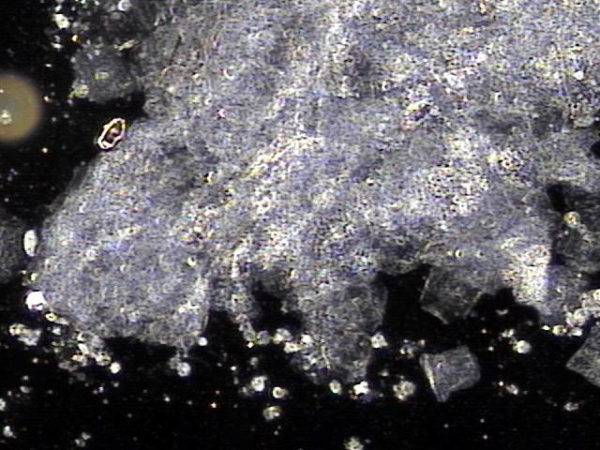
주 3회 전립선의 표적 치료후 전립선관, 사정관과 정관등에 막혀 있던 치료된 상피세포 덩어리의 현미경학적 자료입니다.
This is a microscopic image of treated epithelial cell clusters that had been obstructing the prostatic ducts, ejaculatory ducts, and vas deferens, following targeted prostate therapy administered three times per week.
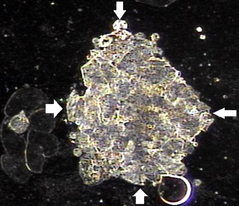
6개월 가량 주 2회 전립선과 정낭, 사정관과 정관등의 표적 치료후 치료된 상피 세포 덩어리의 현미경학적 자료입니다.
This is microscopic evidence of treated epithelial cell clusters following approximately six months of twice-weekly targeted therapy to the prostate, seminal vesicles, ejaculatory ducts, and vas deferens.
주 2회 전립선과 사정관과 정낭 그리고 정관등의 표적 치료후 치료된 현미경학적 자료입니다.
Here is a microscopic image showing materials that were cleared after targeted therapy of the prostate, ejaculatory ducts, seminal vesicles, and vas deferens.
These include shed epithelial cells, inflammatory cells, and some protein-like substances that had been blocking the ducts. Their removal helps restore healthy circulation and improve function in these areas.
전립선과 사정관입구와 사정관 그리고 정낭과 정관의 표적 치료후 치료된 상피세포들의 현미경학적 자료 입니다.
This microscopic image was taken after targeted treatment of the prostate, the entrance of the ejaculatory ducts, the ejaculatory ducts themselves, the seminal vesicles, and the vas deferens.
What you are seeing here is most likely a group of shed epithelial cells along with some inflammatory or fibrous material that had been blocking the ducts. Such blockages can form due to long-standing inflammation or circulation issues.
The good news is that these materials are now being released and cleared after treatment, which is an encouraging sign that healing and better circulation are taking place.
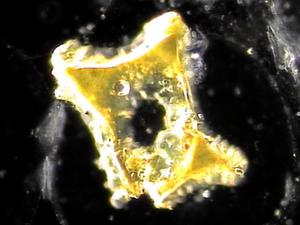
전립선과 사정관입구와 사정관 그리고 정낭과 정관의 표적 치료후 사정관 입구의 결석들이 치료된 현미경학적 자료 입니다.
This microscopic image was taken after targeted treatment of the prostate, the entrance of the ejaculatory ducts, the ejaculatory ducts themselves, the seminal vesicles, and the vas deferens.
The specimen shown here appears to be a calcified deposit or stone-like material (ejaculatory duct calculi). Such structures can form when shed epithelial cells, inflammatory debris, or proteinaceous substances accumulate and harden over time, often as a result of chronic inflammation or impaired circulation in the ducts.
For a more precise interpretation, consultation with international experts in urology, pathology, and andrology would be valuable, as their collective opinions could help confirm whether this represents a true ejaculatory duct stone, calcified tissue, or another type of mineralized deposit.
In practical terms for patients, this image suggests that treatment has helped dislodge and clear these obstructive materials, which is a positive sign for improving ductal flow and overall reproductive health.
This transrectal ultrasound image, taken on the first visit, shows prostate enlargement along with stones at the entrances of both ejaculatory ducts.
The accompanying microscopic findings are from specimens obtained after targeted treatment of the prostate, ejaculatory duct openings, ejaculatory ducts, and vas deferens.
Together, these images illustrate how obstructive materials—such as stones or calcified deposits formed by accumulated epithelial cells, inflammatory debris, or hardened secretions—can be identified and treated.
For patients, this means that the treatment is helping to relieve blockages, improve circulation, and support better prostate and reproductive health.
This approach is quite different from standard surgical options such as laser resection, water jet therapy, TURP, or Rezūm.
Instead of removing or ablating prostate tissue, the focus here is on clearing blockages within the ducts—such as stones, shed epithelial cells, or inflammatory debris—that interfere with circulation and normal function.
The potential benefits are that it may be less invasive, avoid complications from tissue removal, and help restore natural ductal flow. Patients may feel more comfortable, with improved urinary and reproductive health, while maintaining their quality of life.
That said, because this technique is not yet widely studied internationally, it would be valuable to gather more clinical data and expert consensus to better understand its safety, long-term outcomes, and best indications.
Overall, it seems to offer a gentler, restorative approach that could make patients feel happier and more hopeful for a better life without major surgical procedures.
주 2회 전립선과 정낭과 사정관 그리고 정관등의 표적 치료후 치료된 탈락되어 막혀 있던 상피 세포와 프로스타그란딘에 의한 염증 덩어리들의 치료된 현미경학적 사진입니다.
This microscopic image shows the treated area after targeted therapy of the prostate, seminal vesicles, ejaculatory ducts, and vas deferens.
It reveals the removal of old, detached epithelial cells and inflammatory clumps caused by prostaglandins that had been blocking the ducts.
These findings suggest that the blocked passages are now being cleared, helping to restore normal circulation and function.
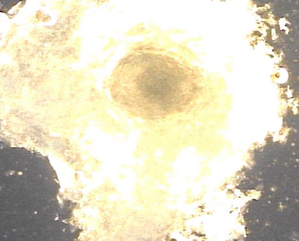
주 2회 전립선과 정낭과 사정관 그리고 정관등의 표적 치료후 치료된 탈락되어 막혀 있던 상피 세포와 결석들의 치료된 현미경학적 자료입니다.
This microscopic image shows the treated materials after targeted therapy of the prostate, seminal vesicles, ejaculatory ducts, and vas deferens.
It reveals the removal of detached epithelial cells and stones that had been blocking the ducts, indicating that the obstructions have been successfully cleared.
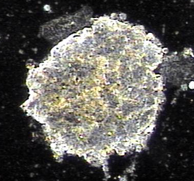
주 2회 전립선과 정낭, 사정관입구와 사정관 그리고 정낭과 정관등의 표적 치료후 치료된 전립선관과 사정관,그리고 정관내의 탈락된 상피 세포와 선상피들의 현미경학적 자료입니다.
These microscopic images show exfoliated epithelial cells and glandular cells from the treated prostatic ducts, ejaculatory ducts, and vas deferens after twice-weekly targeted therapy of the prostate, seminal vesicles, ejaculatory duct openings, and vas deferens.
주 2회 전립선과 정낭과 사정관 그리고 정관등의 표적 치료후 치료된 탈락되어 막혀 있던 상피 세포와 결석들의 치료된 현미경학적 자료입니다.
This microscopic image shows the treated materials after targeted therapy of the prostate, seminal vesicles, ejaculatory ducts, and vas deferens.
It reveals the removal of detached epithelial cells and stones that had been blocking the ducts, indicating that the obstructions have been successfully cleared.
개인의 차가 있을수 있으나
평균적으로 항문에서 전립선까지 거리는 6Cm,
정낭까지의 거리는10Cm, 그리고
정낭의 크기는 가로5Cm,세로2.5Cm,두께1Cm 즉 항문에서 정낭 첨부까진 15Cm 그리고
정관은 좌우 각각 45Cm 항문에서 정관까지 6Cm 이상
사정관까지도 항문에서 6Cm이상 접하여
전립선의 표적치료를 해야하며
사람마다 둔부를 구성하는 대둔근과 중둔근, 소둔근의 크기가 달라
표적치료시 서로 노력하여
만성 전립선염과 전립선 비대증, 전립선의 낭종과 전립선의 결석과 전립선의 암 그리고
정낭의 낭종 고환의 미석증과 만성골반통 증후군등의 반드시
치료되는 전립선의 표적치료를 하러가야합니다.
이따가 뵙겠습니다.
서울가정의학과의원 드림.
While there may be individual variations, on average:
- The distance from the anus to the prostate is 6 cm.
- The distance to the seminal vesicles is 10 cm.
- The size of the seminal vesicles is 5 cm in width, 2.5 cm in height, and 1 cm in thickness, meaning the distance from the anus to the tip of the seminal vesicle is approximately 15 cm.
- The vas deferens extends 45 cm on each side, with at least 6 cm from the anus to the vas deferens and the ejaculatory ducts.
The Importance of Targeted Prostate Treatment
Due to this anatomical structure, targeted prostate treatment is essential. Since the size of the gluteal muscles (gluteus maximus, medius, and minimus) varies among individuals, precise efforts are needed during treatment.
This treatment is crucial for effectively managing and resolving conditions such as:
- Chronic prostatitis
- Benign prostatic hyperplasia (BPH)
- Prostatic cysts and calcifications
- Prostate cancer
- Seminal vesicle cysts
- Testicular microlithiasis
- Chronic pelvic pain syndrome (CPPS)
We highly recommend undergoing targeted prostate treatment to address these issues.
See you soon.
Best regards,
Seoul Family Medicine Clinic
댓글목록
등록된 댓글이 없습니다.


