전립선 자료실
페이지 정보
본문
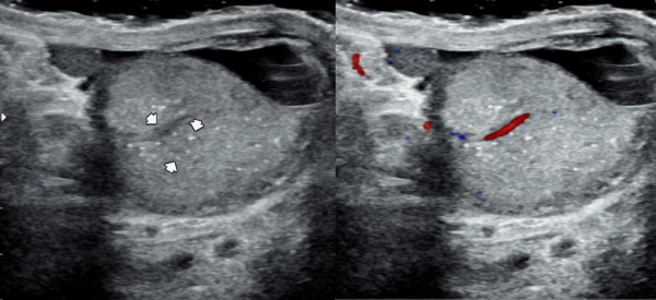
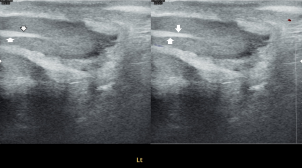
오래전부터 배뇨장애와 빈뇨 및 급박뇨등으로 다른 비뇨기과에서 약물 투여를 했으나 증상의 호전이 없다고 내원 당일 검사한 고환내 다량의 미석증이 관찰된 초음파 사진입니다.
This is an ultrasound image taken on the day of the patient’s visit, who reported long-standing urinary symptoms such as voiding difficulty, frequent urination, and urgency, with no improvement despite medication from other urology clinics. The scan shows a large amount of microcalcifications within the testes.
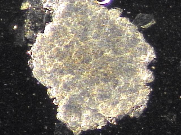
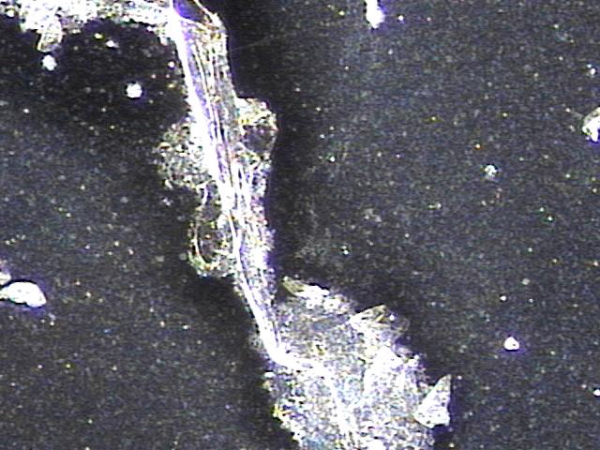
전립선과 정관의 표적 치료후 배출된 상피 세포 덩어리들(주2회 표적치료)
Epithelial cell clusters discharged after targeted treatment of the prostate and vas deferens (twice-weekly targeted treatment).
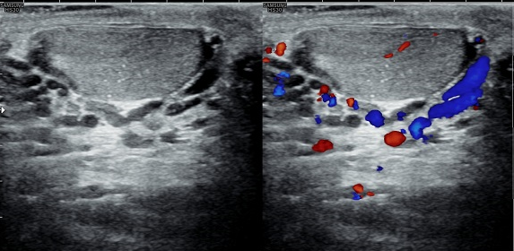
주2회 전립선의 표적 치료후 고환의 미석증이 줄어 들고 있는 경직장 전립선 초음파 검사 사진
Transrectal ultrasound image showing a reduction in testicular microlithiasis after twice-weekly targeted prostate treatment.
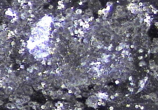
전립선과 정관의 표적 치료후 배출된 상피 세포 덩어리들(주2회 표적치료)
Epithelial cell clusters discharged after targeted treatment of the prostate and vas deferens (twice-weekly targeted treatment).
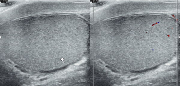
전립선의 표적치료후 고환의 미석증이 없어지고 고환이 커진 경직장 전립선 초음파 자료 입니다.(주2회 표적치료)
This is a transrectal ultrasound image showing the improvement after targeted prostate treatment.
After twice-weekly targeted treatment, testicular microlithiasis disappeared, and the testicles increased in size.
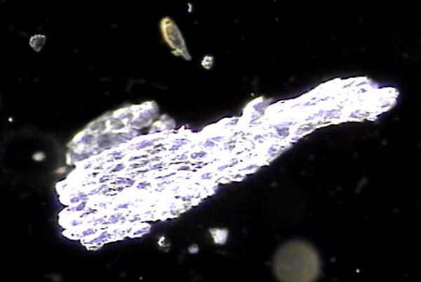
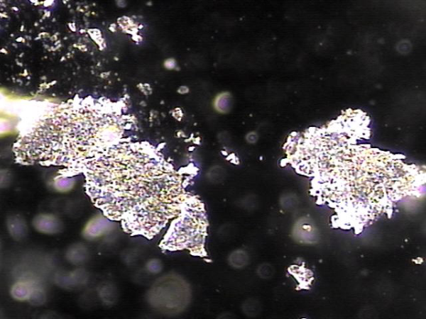
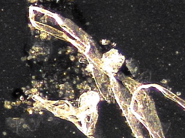
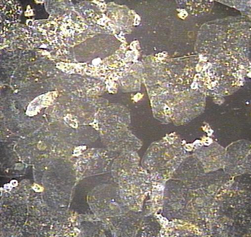
지속적인 전립선의 표적 치료후 치료된 사정관과 정관 그리고 정낭등에 막혀 있던 상피세포 덩어리의 현미경학적 자료입니다.
This is a microscopic image of epithelial cell clusters that had been blocking the ejaculatory ducts, vas deferens, and seminal vesicles, which were cleared following continuous targeted prostate treatment.
The ultrasound image on the left shows the initial examination, where testicular microlithiasis (tiny calcifications) was present.
The ultrasound image on the right is a follow-up study after several months of targeted therapy to the vas deferens, ejaculatory ducts, seminal vesicles, and prostate, twice a week.
The follow-up scan demonstrates that the previously noted microlithiasis has improved, suggesting that the targeted treatment contributed to the restoration of testicular health.
We hope this evidence may help inform colleagues and patients worldwide that such cases of testicular microcalcification, associated with chronic prostatitis or seminal tract obstruction, can improve through this approach.
오랜 세월동안 탈락된 상피세포가 항문 주위에 결석을 형성하여 급박뇨와 야간 빈뇨를 일으키는 배출되지 못한 상피 세포가 전립선과 정관 그리고 사정관등에서 치료된 현미경학적 사진입니다.
This is a microscopic image showing the epithelial cell clusters that had built up over many years, forming small stones around the anal area. These blockages contributed to symptoms like urgency and frequent nighttime urination. With targeted treatment, these cells were successfully cleared from the prostate, vas deferens, and ejaculatory ducts.
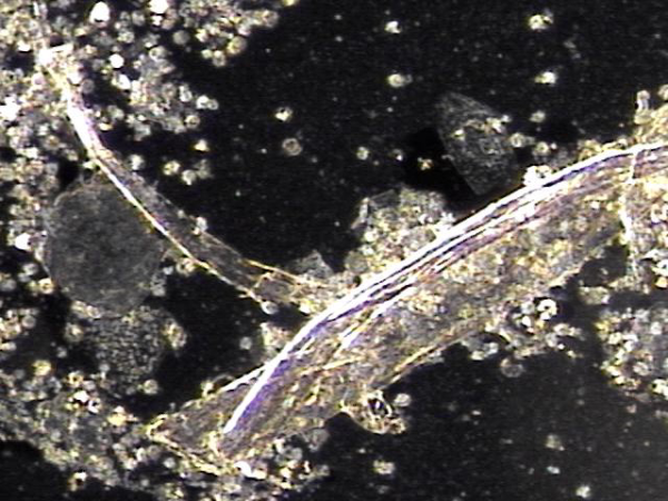
주 2회 전립선과 정낭, 사정관과 정관등의 표적 치료후 막혀 있던 탈락된 상피 세포가 치료된 현미경학적 자료 입니다.
This is a microscopic image taken after twice-weekly targeted treatment of the prostate, seminal vesicles, ejaculatory ducts, and vas deferens. It shows that the shed epithelial cells that had been blocking these areas have been successfully cleared.
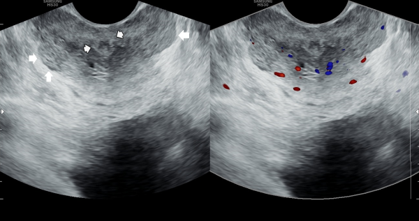
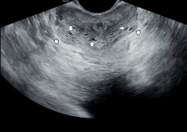
주 2회 전립선의 표적치료중 탈락된 상피 세포가 전립선관을 막아 전립선의 다발성 낭종을 형성하여 전립선 결절과 비대로 급박뇨와 빈뇨등의 증상을 생기게하는 추적 경직장 전립선 초음파 사진입니다.
This follow-up transrectal ultrasound image of the prostate shows that during twice-weekly targeted therapy, shed epithelial cells blocked the prostatic ducts, leading to multiple cysts, nodular enlargement, and symptoms such as urinary urgency and frequency.
주 2회 전립선과 정낭, 사정관과 정관등의 표적 치료후 막혀 있던 탈락된 상피 세포가 치료된 현미경학적 자료 입니다.
This is a microscopic image taken after twice-weekly targeted treatment of the prostate, seminal vesicles, ejaculatory ducts, and vas deferens. It shows that the shed epithelial cells that had been blocking these areas have been successfully cleared.
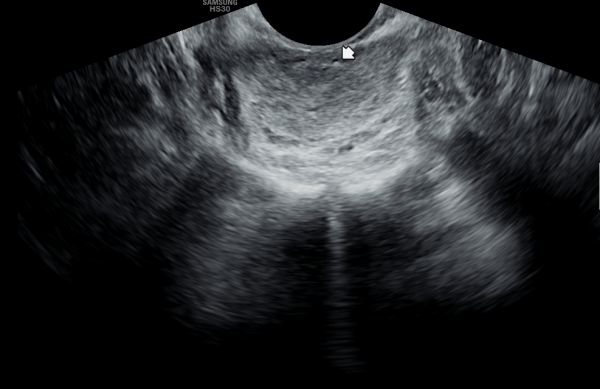
빈뇨와 급박뇨등의 증상을 생기게하는 항문주위의 경직장 전립선 초음파 사진입니다.
This is a transrectal prostate ultrasound image around the anal area, showing changes that can cause symptoms such as frequent urination and urgency.
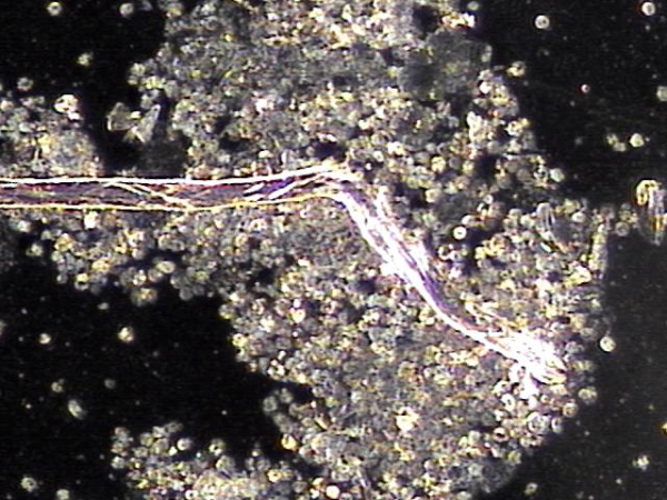
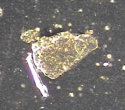
주 2회 전립선과 정낭, 사정관과 정관등의 표적 치료후 막혀 있던 탈락된 상피 세포가 치료된 현미경학적 자료 입니다.
This is a microscopic image taken after twice-weekly targeted treatment of the prostate, seminal vesicles, ejaculatory ducts, and vas deferens. It shows that the shed epithelial cells that had been blocking these areas have been successfully cleared.
주 2회 전립선과 정낭, 사정관과 정관등의 표적 치료후 막혀 있던 탈락된 상피 세포가 치료된 현미경학적 자료 입니다.
This microscopic image shows how the shed epithelial cells that were blocking the prostate, seminal vesicles, ejaculatory ducts, and vas deferens have been cleared after twice-weekly targeted therapy.
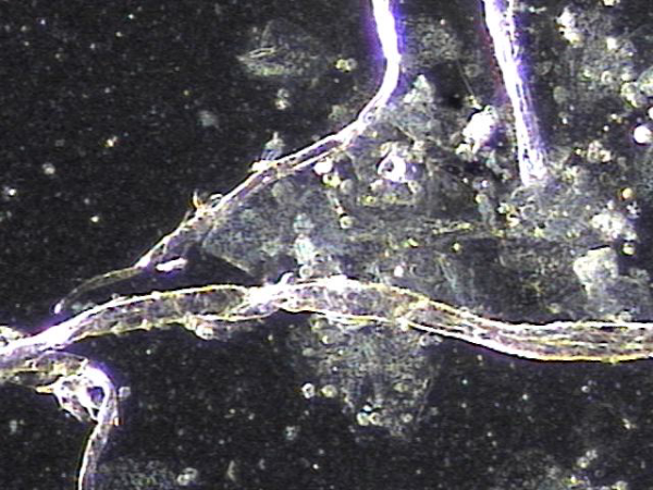
주 2회 전립선과 정낭, 사정관과 정관등의 표적 치료후 막혀 있던 탈락된 상피 세포와 프로스타그란린에 생성된 염증들이 치료된 현미경학적 자료 입니다.
This microscopic image shows how the shed epithelial cells and inflammation caused by prostaglandins, which had been blocking the prostate, seminal vesicles, ejaculatory ducts, and vas deferens, were cleared after twice-weekly targeted therapy.
주 2회 전립선과 사정관, 정낭 그리고 정관등의 표적 치료후 탈락되어 전립선관과 사정관 그리고 정낭과 정관등에 막혀 있던 상피세포가 치료된
현미경학적 자료입니다.
This is a microscopic image taken after targeted treatments, performed twice a week on the prostate, ejaculatory ducts, seminal vesicles, and vas deferens. It shows that the shed epithelial cells, which had been blocking the prostate ducts, ejaculatory ducts, seminal vesicles, and vas deferens, have now been successfully cleared through treatment.
사정관과 정관등의 탈락된 상피 세포가 막혀 정자의 순환 장애를 일으키면 고환의 섬유화를 일으키는 초음파 사진입니다.
This ultrasound image shows how shed epithelial cells in the ejaculatory ducts and vas deferens can block the flow of sperm. When this circulation is disrupted, it may lead to testicular fibrosis (scarring in the testicle).
고환의 섬유화를 일으키는 있는 정관내에 막혀 있던 상피 세포 덩어리가 치료된 현미경학적 자료입니다.
This microscopic image shows clumps of epithelial cells that had been blocking the vas deferens and causing fibrosis of the testis. After targeted therapy, these blockages have been treated.
모든 남성은 누구던지 반드시 기억해야 할 의학적인 지식은 매일 신체의 각 부위에서 충실히 역활을 한 세포는 1~3주내 수명을 다하고 탈락하며
새로운 상피세포가 만들어져 각 장기에 그 역활을 이어가나
전립선과 사정관 그리고 정관의 상피세포는 탈락하여
좁고 긴 배출관(직경 : 0.1~0.3mm)에 쌓여 비대해 지고
낭종과 섬유화와 결석등이 진행하여
여러 증상이 생겨 온갖 광고에 유혹 되기도 하고
잘못된 치료의 길도 갑니다.
꼭 기억 해야할 치료는 오랫동안 쌓인 상피세포 덩어리는
수년간~ 반복 표적치료로 휴유증 없이 치료된다는 자료입니다.^^
전립선관,사정관 그리고 정관등이 막혀 전립선등이 커져
전립선암 수치 지표인 PSA 수치가 높아져 조직검사등
레이져 치료, 전립선 결찰 의 시술, 전립선 절제술등의 진행을 막기위해
반드시 전립선의 표적치료를 하러 가야 합니다. 이따가 뵙겠습니다.
Essential Medical Knowledge Every Man Must Remember
Every man must be aware of an important medical fact: cells in every part of the body fulfill their function and naturally shed after completing their lifespan, typically within 1 to 3 weeks.
New epithelial cells continuously regenerate to maintain the function of each organ. However, in the prostate, ejaculatory ducts, and vas deferens, shed epithelial cells can accumulate in these narrow and long ducts (diameter: 0.1–0.3mm), leading to blockage, enlargement, cyst formation, fibrosis, and calcification.
As a result, various symptoms arise, often leading individuals to fall for misleading advertisements or ineffective treatments.
The Key to Proper Treatment
It is crucial to remember that long-accumulated epithelial cell clusters can be effectively treated through repeated targeted treatment over several years, without long-term complications.
If the prostatic ducts, ejaculatory ducts, and vas deferens become blocked, the prostate enlarges, and PSA levels (a marker for prostate cancer) rise, prompting unnecessary procedures such as biopsies, laser treatments, prostate lift procedures, or even prostatectomy.
To prevent this progression, targeted prostate treatment is essential.
See you soon.
- 이전글전립선자료실 25.03.13
댓글목록
등록된 댓글이 없습니다.


